Posts

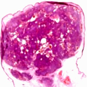
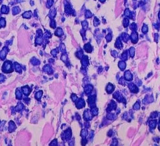
Answer of Dermatopathology Case 100

Retiform Hemangioendothelioma Visit: Dermatopathology Site Visit: Pathology of Hemangioendothelioma Abstract: Retiform haemangioendothelioma: a case report.Ann Pathol. 2009;29(6):491-4. Retiform haemangioendothelioma is a locally aggressive, very rarely metastasizing vascular lesion. Histologically, it is characterized by distinctive arborizing blood vessels resembling "rete testis" and lined by endothelial cells with characteristic hobnail morphology. We present an additional case, in the leg of a 64-year-old patient. We discuss the classification of hemangioendotheliomas. The term hemangioendothelioma should be restricted to vascular tumours of "intermediate malignancy" but has been used to designate tumours with variable histological features and clinical behaviour. Spindle cell hemangio(endothelio)ma is currently regarded as a benign reactive lesion. Kaposiform hemangioendothelioma is potentially lethal due to consumption coagulopathy but no metastasizing case h

Dermatopathology Case 100

Image
Image 1 Image 2 Image 3 Image4 Case 100 A 28 year old male with an indolent brownish plaque on the right lower limb. Diagnosis

Answer of Dermatopathology Case 99

Kaposiform Hemangioendothelioma Visit: Dermatopathology Site Visit: Pathology of Hemangioendothelioma Abstract: Successful treatment of Kasabach-Merritt syndrome arising from kaposiform hemangioendothelioma by systemic corticosteroid therapy and surgery.Int J Clin Oncol. 2011 Sep 28. Kasabach-Merritt syndrome is a rare type of vascular tumor with aggressive behavior in association with thrombocytopenia and consumptive coagulopathy. A standard guideline has not been established to date. A 7-day-old male infant with Kasabach-Merritt syndrome arising from kaposiform hemangioendothelioma was successfully treated with systemic corticosteroid and surgery. Systemic corticosteroid including methylprednisolone was injected intravenously followed by an intralesional injection of compound betamethasone. This approach brought about an excellent response after the first treatment which was maintained long enough to provide us with an opportunity to excise the tumor. Systemic corticosteroid and surg

Dermatopathology Case 99

Image
Image1 Image2 Image3 Image4 Image5 Case 99 A reddish blue nodule on the left upper arm of a 3 year old child. Diagnosis

Answer of Dermatopathology Case 98

Epithelioid Hemangioendothelioma Visit: Dermatopathology site Visit: Pathology of Hemangioendothelioma Abstract Epithelioid hemangioendothelioma presenting in the skin: a clinicopathologic study of eight cases. Am J Dermatopathol.1998 Dec;20(6):541-6. Epithelioid hemangioendothelioma arising in the skin is extremely rare, and the majority of documented cases have developed in association with an underlying bone tumor. We report eight patients with an age range of 29-84 years (mean 53), who presented with primary cutaneous tumors at a variety of sites including the palm, shin, neck, knee, nose, back, and penis with a duration of between 6 and 12 months. Histologically, all eight cases presented as circumscribed nodules with an overlying acanthotic epidermis, three showing striking acrosyringeal proliferation, reminiscent of eccrine syringofibroadenoma. The tumors were composed of an admixture of slightly pleomorphic spindle and epithelioid cells with abundant, sharply defined eosinophil

Dermatopathology Case 98

Image
Image1 Image2 Image3 Image4 Image5 Case 98 A 45 year old woman with an erythematous, soft, painful nodule on the neck. Diagnosis

Answer of Dermatopathology Case 97

Image
Immunohistochemistry findings: CD3 CD4 CD7 CD20 Mycosis Fungoides-associated Follicular Mucinosis: Abstract: Follicular mucinosis: a clinicopathologic, histochemical, immunohistochemical and molecular study comparing the primary benign form and the mycosis fungoides- associated follicular mucinosis.J Cutan Pathol. 2009 Jul 14. Objectives: To determine (i) whether primary (idiopathic) follicular mucinosis (PFM) and lymphoma-associated follicular mucinosis (LAFM) are distinct or related entities and whether there are reliable criteria that allow the two forms to be distinguished, (ii) the histochemical properties and consequently the type of mucin that accumulates in the follicle in PFM and LAFM, and (iii) whether there is any difference between the staining properties of mucin in patients with PFM and LAFM. Methods: Thirty-one patients were divided into two groups. Group 1 comprised 20 patients with no associated mycosis fungoides or Sézary syndrome (PFM) and group 2 was made up of

Dermatopathology Case 97

Image
Image1 Image2 Image3 Image4 Case 97 A 56 year old woman with violaceous follicular nodules and indurated plaques on the face and neck. Diagnosis

Answer of Dermatopathology Case 96

Image
Immunohistochemistry: CD30 positive cells [ CD3 & CD4 are faintly positive ; ALK and EMA -Negative] Primary Cutaneous Anaplastic Large Cell Lymphoma Abstracts: Primary cutaneous anaplastic large-cell lymphoma.Dermatol Online J. 2010 15;16(11):2. Primary cutaneous anaplastic large-cell lymphoma (ALCL) is a form of cutaneous T-cell lymphoma that is characterized by solitary or localized nodules or plaques. Histopathologic features include a diffuse, non-epidermotropic infiltrate with cohesive sheets of large anaplastic CD30+ tumor cells. This entity must be distinguished from systemic ALCL with cutaneous involvement and lymphomatoid papulosis. Treatment modalities include clinical monitoring, radiation therapy, and surgical excision, with systemic chemotherapy reserved for disseminated or extracutaneous disease. CD30(+) anaplastic large cell lymphoma: a review of its histopathologic, genetic, and clinical features. Blood.2000 Dec 1;96(12):3681-95. Anaplastic large cell lymphoma (ALCL

Dermatopathology Case 96

Image
Image1 Image2 Image 3 Image4 Case 96 A 55 year old male with an ulcerated nodule on the back of the left arm. Diagnosis

Answer of Dermatopathology Case 95

Papillary Eccrine Adenoma Visit: Dermatopathology site Visit: Papillary Eccrine Adenoma Abstract: Papillary eccrine adenoma in association with cutaneous horn. Am J Clin Dermatol. 2007;8(3):179-82. Papillary eccrine adenoma (PEA) is an uncommon sweat gland neoplasm that occurs more frequently on the distal extremities of Black women. Clinically, it appears as a small, isolated, firm, dome-shaped cutaneous nodule. We report two unusual cases of PEA that presented clinically as cutaneous horn. Histologically, both cases showed the typical dermal morphology of PEA with overlying epidermal changes suggestive of human papillomavirus (HPV). However, HPV immunoperoxidase staining and polymerase chain reaction for HPV were negative in both cases. Concurrent occurrence of PEA and HPV-like epidermal changes may be coincidental; however, their co-occurrence may also be related to the environment induced by this adnexal tumor. Whether PEA with verrucous epidermal changes has a different clinical b

Dermatopathology Case 95

Image
Image1 Image2 Image3 Image4 Image5 Case 95 A 45 year old male with a firm nodule on the dorsum of his right hand. Diagnosis

Answer of Dermatopathology Case 94

Image
Hypertrophic Lichen Planus Visit: Dermatopathology site [ Related links to clinical images of similar condition: (DermAtlas -JHU.): Image1 ; Image2 ; Image3 ; Image4 ] Abstract: Verrucous squamous cell carcinoma complicating hypertrophic lichen planus : Three case reports and review of the literature. Hautarzt. 2011 Jan;62(1):40-45. Lichen planus is a chronic mucocutaneous T-cell-mediated disease, whose cause is still unknown. The first case of lichen planus that transformed into squamous cell carcinoma was reported in 1903. We present three patients in whom squamous cell carcinomas were identified in chronic lichen planus. The world literature includes at least 91 cases, including our three cases. In an epidemiological study, no significant risk of transformation of cutaneous lichen planus into squamous cell carcinomas was found. In contrast, there is a significantly higher risk of malignant transformation in mucosal lichen planus, so that the WHO had graded mucosal lichen planus a

Dermatopathology Case 94

Image
Image1 Image2 Image3 Image4 Case 94 A 20 year old male with multiple pruritic plaques on the extremities. The lesions have a verrucous appearance. Diagnosis [ Related links to clinical images of similar condition: (DermAtlas -JHU.): Image1 ; Image2 ; Image3 ; Image4 ]

Answer of Dermatopathology Case 93

Image
Giemsa stain Toluidine Blue stain Urticaria Pigmentosa Visit: Dermatopathology site Related links to clinical images of the condition: (DermAtlas -JHU.): Image1 ; Image2 ; Image3 ; Image4 . Abstract: Diagnostic criteria and classification of mastocytosis: a consensus proposal. Leuk Res. 2001 Jul;25(7):603-25. The term 'mastocytosis' denotes a heterogeneous group of disorders characterized by abnormal growth and accumulation of mast cells (MC) in one or more organ systems. Over the last 20 years, there has been an evolution in accepted classification systems for this disease. In light of such developments and novel useful markers, it seems appropriate now to re-evaluate and update the classification of mastocytosis. Here, we propose criteria to delineate categories of mastocytosis together with an updated consensus classification system. In this proposal, the diagnosis cutaneous mastocytosis (CM) is based on typical clinical and histological skin lesions and absence of def

Dermatopathology Case 93

Image
Image1 Image2 Image3 Image4 Image5 Case 93 A 2 year old child with multiple reddish brown macules on the trunk. Diagnosis and special stains : [ Related links to clinical images of similar condition: (DermAtlas -JHU.): Image1 ; Image2 ; Image3 ; Image4 .]

Answer of Dermatopathology Case 92

Subcorneal Pustular Dermatosis (Sneddon-Wilkinson disease) Visit: Dermatopathology site Abstract: Subcorneal pustular dermatosis: 50 years on.Clin Exp Dermatol. 2008 May;33(3):229-33. Epub 2008 Mar 18. We review the key developments in our understanding of subcorneal pustular dermatosis (SCPD, also known as Sneddon-Wilkinson disease) over the past 50 years. SCPD is a rare, chronic, sterile pustular eruption that was first described by Sneddon and Wilkinson in 1956. The primary lesions are pea-sized pustules classically described as half-pustular, half-clear flaccid fluid blisters. Histologically the salient feature is a subcorneal accumulation of neutrophils, suggesting the presence of chemoattractants such as tumour necrosis factor (TNF)alpha in the uppermost epidermis. However, to date its exact pathophysiology is unknown. Cases in association with pyoderma gangrenosum, benign monoclonal IgA gammopathy and multiple myeloma are well documented. There are anecdotal reports of SCPD asso

Dermatopathology Case 92

Image
Image1 Image2 Image3 Image4 Case 92 A 52 year old woman, with pustules about 4 mm in diameter, on the neck. Diagnosis

Answer of Dermatopathology Case 91

Proliferating Trichilemmal Cyst (Pilar Tumour) Visit: Dermatopathology site Visit: Proliferating Trichilemmal Cyst (Pilar Tumour) Abstract: Proliferating trichilemmal tumors: a review of the literature. Dermatol Surg. 2007 Sep;33(9):1102-8. BACKGROUND: Proliferating trichilemmal tumors (PTTs) are uncommon lesions whose histologic hallmark is the presence of trichilemmal keratinization. PTT is thought to originate from the trichilemmal cyst (TC) and have the potential for malignant transformation, at which point it is termed a malignant proliferating trichilemmal tumor (MPTT). These lesions may cause considerable morbidity and even mortality, and recurrence after simple local excision is common. OBJECTIVE: The objective was to review the clinical presentation, histopathologic characteristics, malignant potential, and treatment options for PTT. METHODS: The English literature was reviewed regarding PTT, TC, and MPTT. RESULTS: PTT generally presents as a subepidermal tumor on the scalp in