Posts

Showing posts from December, 2009

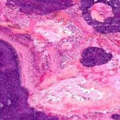
Answer of Dermatopathology Case 28

Molluscum Contagiosum Visit: Molluscum Contagiosum Visit: Dermatopathology site Visit: Infectious Disease Online Abstract: Mollusca contagiosa. From paediatric dermatology to sexually transmitted infection.Hautarzt. 2009 Jun;60(6):472-6. Molluscum contagiosum (MC) is a common cutaneous infection caused by the molluscipox virus (MCV) and can affect both children and adults. Molluscum contagiosum is relatively frequent in children aged 1-5 years old and can be localized almost anywhere on the body, but in adults it is regarded as a sexually transmitted infection (STI). MCV can be transmitted directly from person to person or by autoinoculation. MC in adults characteristically involves the genital area but extragenital appearance can be more typically seen in patients with immunosuppressive conditions, especially in HIV/AIDS. The onset of MC inHIV-positive individuals can be regarded as a part of the immune reconstitution inflammatory syndrome (IRIS). MC probably affects both sexes equall

Dermatopathology Case 28

Image
Image1 Image2 Image3 Case 28 A 17 year old boy with a pearly white papule on the forehead. Answer

Answer of Dermatopathology Case 27

Sebaceoma Visit: Pathology of Sebaceoma Visit: Dermatopathology site Visit: Sebaceous Tumours and Tumour-like Lesions Abstract: Sebaceous lesions and their associated syndromes: part I. J Am Acad Dermatol. 2009 Oct;61(4):549-60; quiz 561-2. Sebaceous neoplasms have long been a source of confusion to dermatologists and pathologists alike. Disagreements regarding nomenclature, classification, and management have been longstanding. Sebaceous lesions represent a broad spectrum of interesting entities that range from hamartomas, hyperplasias, and benign tumors to highly malignant neoplasms. This article discusses the clinical and pathologic features of sebaceous hyperplasia, nevus sebaceous of Jadassohn, sebaceous adenoma, seboacanthoma, sebaceous epithelioma, sebaceoma, mantleoma, basal cell carcinoma with sebaceous differentiation, sebomatricoma(sebomatrixoma), and sebaceous carcinoma. Controversies regarding these lesionswill be explored, and any relationship with Muir-Torre syndrome wil

Dermatopathology Case 27

Image
Image1 Image2 Image3 Image4 Case 27 A 40 year old male with a solitary nodule (2 cm in diameter) on the left side of the face. Answer

Answer of Dermatopathology Case 26

Glomangioma Visit: Pathology of Glomus Tumour Visit: Dermatopathology site Visit: Vascular Tumor Abstract: Sclerotic Glomus Tumor. Am J Dermatopathol. 2009 Oct 30. We report an unusual histopathological variant of a glomus tumor that arose in a peculiar topographic site, a sclerotic glomus tumor. Unlike conventional glomus tumors or glomangiomas that have a loose fibrous stroma with variable hyaline and myxoid changes, the case reported herein had a diffuse, hyalinized, sclerotic stroma. A further difference was that the majority of glomus tumors and glomangiomas occur in the subungual area, trunk, or extremities, whereas the present tumor occurred on the ear. Due to the peculiar histological features and location, other tumors were considered in the differential diagnosis to include Merkel cell carcinoma, primitive neuroectodermal tumor, and small cell melanoma. This article illustrates a unique variant of a glomus tumor, which to our knowledge has not been previously described. Local

Dermatopathology Case 26

Image
Image1 Image2 Image3 Image4 Case 26 A 45 year old female with a painful, bluish nodule on the left upper arm. Answer

Answer of Dermatopathology Case 25

Clear Cell Acanthoma Visit: Pathology of Clear Cell Acanthoma Visit: Dermatopathology site Abstract: Comparing in vivo reflectance confocal microscopy, dermoscopy, and histology ofclear-cell acanthoma . Dermatol Surg. 2009 Jun;35(6):952-9. BACKGROUND: Clear cell acanthoma (CCA) is a rare, benign neoplasm of unknown etiology, whose dermoscopic and histological features have been previously described. Usually, CCA can be diagnosed by clinical and dermoscopic examination. In some cases, diagnosis remains uncertain, and histological examination is needed. The aim of this paper was to describe the features of reflectance confocal microscopy (RCM) in diagnosing CCA, compare them with findings on dermoscopy and histology, and evaluate their possible usefulness in CCA evaluation. PATIENTS AND METHODS: Five lesions diagnosed clinically as CCA were imaged using dermoscopy and RCM. All lesions were surgically excised to confirm the diagnosis and compare the morphological attributes under light mi

Dermatopathology Case 25

Image
Image1 Image2 Image3 Image4 Image5 Case 25 A 34 year old male with a slow growing reddish-brown papule on the left lower leg. Answer

Answer of Dermatopathology Case 24

Poroma Visit: Pathology of Poroma Visit: Benign Sweat Gland Tumours Visit: Dermatopathology site Abstract: Poromas and porokeratosis in a patient treated for solid-organ and haematological malignancies. Clin Exp Dermatol. 2009 Dec 2. We describe a patient with previous solid-organ (testicular, oesophageal) and haematological (acute myeloid leukaemia) malignancies, in whom chronic cutaneous graft-versus-host disease was complicated by poromas and porokeratosis. Chemotherapy, total body irradiation, longstanding immunosuppression and ultraviolet radiation may all have played a part in the pathogenesis of the skin tumours. Poroma of the hip and buttock.Dermatol Online J. 2009 May 15;15(5):10. Poroma is a benign adnexal skin tumor seen in middle aged individuals with no sex predilection. The acral sites are the most commonly affected regions. Hip or buttock as a location of origin has rarely been reported. We report two cases of poroma, one located on the hip of a 75-year-old man and the o

Dermatopathology Case 24

Image
Image1 Image2 Image3 Image4 Case 24 A 62 year old male with a greyish brown nodule on the left sole. The nodule is 1.5 cm in diameter. Patient has a history of oesophageal carcinoma. Answer

Answer of Dermatopathology Case 23

Pilomatrixoma (Pilomatricoma ; Calcifying epithelioma of Malherbe) Visit: Pathology of Pilomatrixoma Visit: Dermatopathology site Abstract: Fine needle aspiration cytology of pilomatrixoma and differential diagnoses.Acta Cytol. 2009 Nov-Dec;53(6):683-8. OBJECTIVE: To review the cytologic features of pilomatrixoma to allow a definitive diagnosis at the time of aspiration and correct patient management. STUDY DESIGN: Three patients each presented with a mass lesion and on fine needle aspiration were diagnosed as either having pilomatrixoma or a benign skin adnexal tumor. The diagnosis was confirmed on histology. A retrospective analysis of the cytology was performed. RESULT: The most consistent and predominant features for diagnosis were basaloid cells, anucleate squamous cells and calcification in an inflammatory background, often with giant cells. Only focal evidence of true shadow or "ghost" cells was seen in one case on the Diff-Quik smear. Therefore, lack of shadow cells d

Dermatopathology Case 23

Image
Image1 Image2 Image3 Image4 Case 23 A 5 year old boy with a solitary firm nodule on the cheek. Answer

Answer of Dermatopathology Case 22

Fibroepithelioma of Pinkus Visit: Basal Cell Carcinoma Visit: Dermatopathology site Abstract: Variations in clinical presentation of basal cell carcinoma. Acta Clin Croat. 2008 Mar;47(1):25-30. Basal cell carcinoma (basalioma, BCC) is the most common skin cancer and the mostcommon human malignancy in general, with a continuously increasing incidence. In most cases, BCC develops on chronically sun-exposed skin in elderly people, most commonly in the head and neck region. Besides chronic UV radiation, other risk factors for the development of BCC include sun bed use, family history of skin cancer, skin type 1 and 2, a tendency to freckle in childhood, immunosuppression, previous radiotherapy, and chronic exposure to certain toxic substances such as inorganic arsenic. There are numerous variations in clinical presentation of BCC, such as nodular BCC, ulcerating BCC, pigmented BCC, sclerosing BCC, superficialBCC, and fibroepithelioma of Pinkus. Each varies in terms of clinicalpresentation,

Dermatopathology Case 22

Image
Image1 Image2 Image3 Image4 Case 22 A 54 year old male presents with a solitary, pedunculated on the left upper extremity. The lesion is reddish brown in colour and 1.5 cm in diameter. Answer

Answer of Dermatopathology Case 21

Bowen's Disease Visit: Pathology of Bowen's Disease Visit: Dermatopathology site Abstract: Bowen's disease of the nipple in a young man with AIDS: a case report.Clin Breast Cancer. 2009 Feb;9(1):53-5. Bowen's disease, or squamous cell carcinoma in situ (SCCIS) of the skin, is a malignant neoplasm restricted to the epidermis, without evidence of dermal invasion. It usually develops in sun-exposed area of skin, but other sites can also be affected. Bowen's disease of the nipple is extremely rare and has thus far been reported only in women. We present the case of Bowen's disease of the nipple in an HIV-positive male patient who presented with a scaly lesion on nipple for one year. He also had past genital infection with human papillomavirus, but he was found to be negative for high-risk subtypes. Biopsy of the lesion revealed SCCIS of the nipple areola complex, with extension into the underlying lactiferous ducts of the breast. There was no evidence of invasive ca

Dermatopathology Case 21

Image
Image1 Image2 Image3 Image4 Case 21 A 65 year old male with a solitary, asyptomatic, slowly growing, erythematous, well-demarcated scaly patch on the left side of the neck, 4 cm in diameter. Answer

Dermatopathology Case Index: Case 1 to Case 20

Dermatopathology Case Index : Case 1 = Cutaneous Leiomyosarcoma Case 2 = Sebaceous adenoma in a patient with Muir-Torre syndrome Case 3 = Large Cell Acanthoma Case 4 = Eccrine Porocarcinoma Case 5 = Colloid Milium Case 6 = Primary Cutaneous Apocrine Carcinoma Case 7 = Primary Cutaneous Adenoid Cystic Carcinoma Case 8 = Cutaneous Lymphadenoma Case 9 = Eccrine Spiradenoma Case 10 = Primary Cutaneous Actinomycosis Case 11 = Aggressive Digital Papillary Adenocarcinoma Case 12 = Trichoepithelioma Case 13 = Trichofolliculoma Case 14 = Warty Dyskeratoma Case 15 = Cylindroma Case 16 = Trichilemmoma Case 17 = Fordyce's Spot (Ectopic sebaceous glands) Case 18 = Endometriosis Case 19 = Spiradenocarcinoma (Malignant Eccrine Spiradenoma) Case 20 = Dermatophyte Infection

Answer of Dermatopathology Case 20

Dermatophyte Infection Visit: Dermatophytosis Visit: Dermatopathology site Abstracts: Dermatophyte infections.Am Fam Physician. 2003 Jan 1;67(1):101-8. Dermatophytes are fungi that require keratin for growth. These fungi can cause superficial infections of the skin, hair, and nails. Dermatophytes are spread by direct contact from other people (anthropophilic organisms), animals (zoophilicorganisms), and soil (geophilic organisms), as well as indirectly from fomites. Dermatophyte infections can be readily diagnosed based on the history, physical examination, and potassium hydroxide (KOH) microscopy. Diagnosis occasionally requires Wood's lamp examination and fungal culture or histologic examination.Topical therapy is used for most dermatophyte infections. Cure rates are higher and treatment courses are shorter with topical fungicidal allylamines than with fungistatic azoles. Oral therapy is preferred for tinea capitis, tinea barbae, and onychomycosis. Orally administered griseofulv

Dermatopathology Case 20

Image
Image1 Image2 Image3 Case 20 24 year old male. Skin biopsy for diagnosis. Answer

Answer of Dermatopathology Case 19

Spiradenocarcinoma (Malignant Eccrine Spiradenoma) Visit: Pathology of Spiradenocarcinoma Visit: Dermatopathology site Abstract: Spiradenocarcinoma arising from a spiradenocylindroma: unusual case withlymphoepithelioma-like areas. J Cutan Med Surg. 2009 Jul-Aug;13(4):215-20. BACKGROUND: Hybrid skin adnexal tumors are common, and spiradenocylindroma is well described. OBJECTIVE: However, malignant transformation in this setting isinfrequent, especially resemblance to lymphoepithelioma-like carcinoma of skin,which is not associated with Epstein-Barr virus. METHODS: A 65-year-old female presented with ataxia and a skin nodule composed of a hybrid adnexal tumor(spiradenoma and cylindroma) that transitioned into an undifferentiated carcinoma with attendant lymphocytes and plasma cells. There was widespread disseminationof the undifferentiated component to regional neck lymph nodes. RESULTS: The undifferentiated component resembled a lymphoepithelioma-like carcinoma and showed focal ev

Dermatopathology Case 19

Image
Image1 Image2 Image3 Image4 Case 19 A 65 year old woman with a long standing nodule on the scalp. The asymptomatic nodule was present for last 20 years. Since last two months the nodule became tender and rapidly enlarged in size. The lesion is now 7 cm in diameter. Answer

Answer of Dermatopathology Case 18

Endometriosis Visit: Dermatopathology site Note: Cutaneous endometriosis indicates that scar tissue is particularly susceptible to endometrial implantation. Abdominal wall endometriosis is usually associated with incisional abdominal scars and occurs most commonly following a Caesarean section. It has been suggested that the umbilicus acts as a physiologic scar with a predisposition for developing endometriosis. Umbilical endometriosis has been well described in the literature, occurring either spontaneously or following laparoscopic procedures in which a trocar was placed through the umbilicus. Abstract: Cutaneous endometriosis.Singapore Med J. 2008 Sep;49(9):704-9. INTRODUCTION: Cutaneous or subcutaneous endometriosis is a rare entity that is anoften overlooked because of chronic abdominal pain. METHODS: We reviewed the ten cases of cutaneous endometriosis that presented to this hospital over a seven-year period. RESULTS: The mean age of patients at presentation was 36.3 years. All

Dermatopathology Case 18

Image
Image1 Image2 Image3 Image4 Case 18 A 28 year old woman with a history of Caesarian section 2 years ago. There was a low transverse incision scar. A bluish- gray tender mass developed on the scar. The lesion is 3 cm in diameter. Answer