Posts

Showing posts from January, 2014

Answer of Dermatopathology Case 124

Diagnosis: Pancreatic Panniculitis Visit: Pancreatic Panniculitis

Dermatopathology Case 124

Image
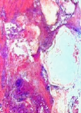
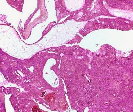
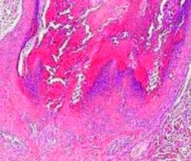
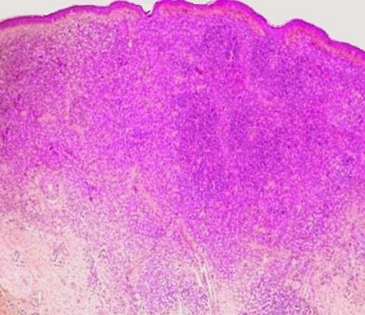
Image1 (Low power)                                             Image2  (Low power)             Image3 (High power)         Image4 (High power)   Case 124: A 45 year old man with painful reddish brown nodule on the left thigh. Patient also has a history of acute pancreatitis.    Spot Diagnosis:

Answer of Dermatopathology Case 123

Answer of Dermatopathology Case 123 : Leukocytoclastic Vasculitis   Visit: Vasculopathic Reaction Pattern

Dermatopathology Case 123

Image
Image1 Image2 Image3 Image4 Case 123   A 35 year old male with an erythematous macule on the left lower leg. Answer

Answer of Dermatopathology Case 122

Diagnosis: Nummular Dermatitis Visit: Spongiotic Reaction Pattern

Dermatopathology Case 122

Image
Image1   Image2 Image3   Image 4 Case 122 Coin shaped multiple patches. The surface is crusted. The lesion is located on the left upper arm of a 50 year old male. Answer

Answer of Dermatopathology Case 121

Answer:  Aggressive digital papillary adenocarcinoma (Digital  Papillary Adenocarcinoma)  For further information: Visit: Aggressive digital papillary adenocarcinoma

Dermatopathology Case 121

Image
Image1 Image2 Image3 Image4 Image5 Image6 Case 121:   A  68 year old male with a tan-gray, solitary cystic nodule, 1.5cm in diameter on the left middle finger. Answer:  

Answer of Dermatopathology Case 120

Answer:   Warty Dyskeratoma For further information:  Visit-  Warty Dyskeratoma

Dermatopathology Case 120

Image
Image1 Image2 Image3 Image4 Image5 Case 120 -  A 50 year old man with a solitary nodule on the forehead. Answer:

Answer of Dermatopathology Case 119

Answer:  Bowen's Disease For further information:  Visit:   Bowen's Disease

Dermatopathology Case 119

Image
Image1 Image2 Image3 Image4 Case 119   A 65 year old male, with a red, scaly patch on the left upper arm. Answer:

Answer of Dermatopathology Case 118

Answer: Halo Naevus ( Sutton Nevus) For more information visit: Halo Naevus (Nevus with halo ; Sutton Nevus)

Dermatopathology Case 118

Image
Image1 Image2 Image3 Image4 Case 118  A reddish brown papule on the back of a 14 year old boy. The lesion is surrounded by a white depigmented ring. Answer :  

Answer of Dermatopathology Case 117

Answer : Cherry Hemangioma For further information visit:  Cherry Hemangioma (Senile Angioma)                

Dermatopathology Case 117

Image
Image1 Image2 Image3 Image4 Case 117    A red papule on the right upper arm of a 62 year old male.  Spot Diagnosis : Answer