Posts

Showing posts from February, 2011

Answer of Dermatopathology Case 97

Image
Immunohistochemistry findings: CD3 CD4 CD7 CD20 Mycosis Fungoides-associated Follicular Mucinosis: Abstract: Follicular mucinosis: a clinicopathologic, histochemical, immunohistochemical and molecular study comparing the primary benign form and the mycosis fungoides- associated follicular mucinosis.J Cutan Pathol. 2009 Jul 14. Objectives: To determine (i) whether primary (idiopathic) follicular mucinosis (PFM) and lymphoma-associated follicular mucinosis (LAFM) are distinct or related entities and whether there are reliable criteria that allow the two forms to be distinguished, (ii) the histochemical properties and consequently the type of mucin that accumulates in the follicle in PFM and LAFM, and (iii) whether there is any difference between the staining properties of mucin in patients with PFM and LAFM. Methods: Thirty-one patients were divided into two groups. Group 1 comprised 20 patients with no associated mycosis fungoides or Sézary syndrome (PFM) and group 2 was made up of

Dermatopathology Case 97

Image
Image1 Image2 Image3 Image4 Case 97 A 56 year old woman with violaceous follicular nodules and indurated plaques on the face and neck. Diagnosis

Answer of Dermatopathology Case 96

Image
Immunohistochemistry: CD30 positive cells [ CD3 & CD4 are faintly positive ; ALK and EMA -Negative] Primary Cutaneous Anaplastic Large Cell Lymphoma Abstracts: Primary cutaneous anaplastic large-cell lymphoma.Dermatol Online J. 2010 15;16(11):2. Primary cutaneous anaplastic large-cell lymphoma (ALCL) is a form of cutaneous T-cell lymphoma that is characterized by solitary or localized nodules or plaques. Histopathologic features include a diffuse, non-epidermotropic infiltrate with cohesive sheets of large anaplastic CD30+ tumor cells. This entity must be distinguished from systemic ALCL with cutaneous involvement and lymphomatoid papulosis. Treatment modalities include clinical monitoring, radiation therapy, and surgical excision, with systemic chemotherapy reserved for disseminated or extracutaneous disease. CD30(+) anaplastic large cell lymphoma: a review of its histopathologic, genetic, and clinical features. Blood.2000 Dec 1;96(12):3681-95. Anaplastic large cell lymphoma (ALCL

Dermatopathology Case 96

Image
Image1 Image2 Image 3 Image4 Case 96 A 55 year old male with an ulcerated nodule on the back of the left arm. Diagnosis

Answer of Dermatopathology Case 95

Papillary Eccrine Adenoma Visit: Dermatopathology site Visit: Papillary Eccrine Adenoma Abstract: Papillary eccrine adenoma in association with cutaneous horn. Am J Clin Dermatol. 2007;8(3):179-82. Papillary eccrine adenoma (PEA) is an uncommon sweat gland neoplasm that occurs more frequently on the distal extremities of Black women. Clinically, it appears as a small, isolated, firm, dome-shaped cutaneous nodule. We report two unusual cases of PEA that presented clinically as cutaneous horn. Histologically, both cases showed the typical dermal morphology of PEA with overlying epidermal changes suggestive of human papillomavirus (HPV). However, HPV immunoperoxidase staining and polymerase chain reaction for HPV were negative in both cases. Concurrent occurrence of PEA and HPV-like epidermal changes may be coincidental; however, their co-occurrence may also be related to the environment induced by this adnexal tumor. Whether PEA with verrucous epidermal changes has a different clinical b

Dermatopathology Case 95

Image
Image1 Image2 Image3 Image4 Image5 Case 95 A 45 year old male with a firm nodule on the dorsum of his right hand. Diagnosis

Answer of Dermatopathology Case 94

Image
Hypertrophic Lichen Planus Visit: Dermatopathology site [ Related links to clinical images of similar condition: (DermAtlas -JHU.): Image1 ; Image2 ; Image3 ; Image4 ] Abstract: Verrucous squamous cell carcinoma complicating hypertrophic lichen planus : Three case reports and review of the literature. Hautarzt. 2011 Jan;62(1):40-45. Lichen planus is a chronic mucocutaneous T-cell-mediated disease, whose cause is still unknown. The first case of lichen planus that transformed into squamous cell carcinoma was reported in 1903. We present three patients in whom squamous cell carcinomas were identified in chronic lichen planus. The world literature includes at least 91 cases, including our three cases. In an epidemiological study, no significant risk of transformation of cutaneous lichen planus into squamous cell carcinomas was found. In contrast, there is a significantly higher risk of malignant transformation in mucosal lichen planus, so that the WHO had graded mucosal lichen planus a

Dermatopathology Case 94

Image
Image1 Image2 Image3 Image4 Case 94 A 20 year old male with multiple pruritic plaques on the extremities. The lesions have a verrucous appearance. Diagnosis [ Related links to clinical images of similar condition: (DermAtlas -JHU.): Image1 ; Image2 ; Image3 ; Image4 ]

Answer of Dermatopathology Case 93

Image
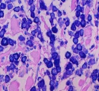
Giemsa stain Toluidine Blue stain Urticaria Pigmentosa Visit: Dermatopathology site Related links to clinical images of the condition: (DermAtlas -JHU.): Image1 ; Image2 ; Image3 ; Image4 . Abstract: Diagnostic criteria and classification of mastocytosis: a consensus proposal. Leuk Res. 2001 Jul;25(7):603-25. The term 'mastocytosis' denotes a heterogeneous group of disorders characterized by abnormal growth and accumulation of mast cells (MC) in one or more organ systems. Over the last 20 years, there has been an evolution in accepted classification systems for this disease. In light of such developments and novel useful markers, it seems appropriate now to re-evaluate and update the classification of mastocytosis. Here, we propose criteria to delineate categories of mastocytosis together with an updated consensus classification system. In this proposal, the diagnosis cutaneous mastocytosis (CM) is based on typical clinical and histological skin lesions and absence of def

Dermatopathology Case 93

Image
Image1 Image2 Image3 Image4 Image5 Case 93 A 2 year old child with multiple reddish brown macules on the trunk. Diagnosis and special stains : [ Related links to clinical images of similar condition: (DermAtlas -JHU.): Image1 ; Image2 ; Image3 ; Image4 .]

Answer of Dermatopathology Case 92

Subcorneal Pustular Dermatosis (Sneddon-Wilkinson disease) Visit: Dermatopathology site Abstract: Subcorneal pustular dermatosis: 50 years on.Clin Exp Dermatol. 2008 May;33(3):229-33. Epub 2008 Mar 18. We review the key developments in our understanding of subcorneal pustular dermatosis (SCPD, also known as Sneddon-Wilkinson disease) over the past 50 years. SCPD is a rare, chronic, sterile pustular eruption that was first described by Sneddon and Wilkinson in 1956. The primary lesions are pea-sized pustules classically described as half-pustular, half-clear flaccid fluid blisters. Histologically the salient feature is a subcorneal accumulation of neutrophils, suggesting the presence of chemoattractants such as tumour necrosis factor (TNF)alpha in the uppermost epidermis. However, to date its exact pathophysiology is unknown. Cases in association with pyoderma gangrenosum, benign monoclonal IgA gammopathy and multiple myeloma are well documented. There are anecdotal reports of SCPD asso

Dermatopathology Case 92

Image
Image1 Image2 Image3 Image4 Case 92 A 52 year old woman, with pustules about 4 mm in diameter, on the neck. Diagnosis

Answer of Dermatopathology Case 91

Proliferating Trichilemmal Cyst (Pilar Tumour) Visit: Dermatopathology site Visit: Proliferating Trichilemmal Cyst (Pilar Tumour) Abstract: Proliferating trichilemmal tumors: a review of the literature. Dermatol Surg. 2007 Sep;33(9):1102-8. BACKGROUND: Proliferating trichilemmal tumors (PTTs) are uncommon lesions whose histologic hallmark is the presence of trichilemmal keratinization. PTT is thought to originate from the trichilemmal cyst (TC) and have the potential for malignant transformation, at which point it is termed a malignant proliferating trichilemmal tumor (MPTT). These lesions may cause considerable morbidity and even mortality, and recurrence after simple local excision is common. OBJECTIVE: The objective was to review the clinical presentation, histopathologic characteristics, malignant potential, and treatment options for PTT. METHODS: The English literature was reviewed regarding PTT, TC, and MPTT. RESULTS: PTT generally presents as a subepidermal tumor on the scalp in

Dermatopathology Case 91

Image
Image1 Image2 Image3 Case 91 A nodule on the scalp of a 62 year old male. Diagnosis

Answer of Dermatopathology Case 90

Lichen Simplex Chronicus Visit: Dermatopathology site Abstract: Lichen simplex chronicus of the anal region and its differential diagnoses. A case series.Hautarzt.2009 Nov;60(11):907-12. Lichen simplex chronicus (LSC) of the anal region is characterized by massive pruritus, constant itching and a chronic course. Histology is notable for a pseudoepitheliomatous hyperplasia. Correct diagnosis as well as therapy of anal LSC sometimes is difficult. Differential diagnostic considerations include verrucous lichen planus and squamous cell carcinoma. We present three cases and then summarize pathogenesis, diagnostics, differential diagnoses and therapeutic options for lichen simplex chronicus of the anal region. Clinical and histopathological findings of 'psoriatic neurodermatitis' and of typical lichen simplex chronicus. J Eur Acad Dermatol Venereol.2007 Jul;21(6):811-7. BACKGROUND: We have seen several patients with itchy lichenified plaques located bilaterally on the elbows and/or

Dermatopathology Case 90

Image
Image1 Image2 Image3 Image4 Case 90 A 36 year old female with scaly, thickened plaque on the extensor surface of left forearm. There is intermittent severe pruritis. Scratching of the area provides temporary relief. Diagnosis

Answer of Dermatopathology Case 89

Digital Mucous Cyst Visit: Dermatopathology site Abstracts: Digital mucous cysts. J Cutan Med Surg. 2010 Sep-Oct;14(5):199-206 BACKGROUND: Digital mucous cyst (DMC) is a common benign condition, but consensus has not been reached regarding its pathogenesis and treatment. OBJECTIVE: This review provides a concise overview of DMCs. METHODS: The review presents the literature pertaining to the etiology, pathogenesis, classification, clinical features, epidemiology, differential diagnoses, diagnosis, and management of DMCs. RESULTS: DMCs have a predilection for middle-aged patients, a good prognosis, and a high recurrence rate. DMCs may occur in one of three locations on the distal digit. They arise owing to a metaplastic or degenerative process. Preexisting osteoarthritis is common and may be an etiologic factor in patients with DMCs. A number of conservative and surgical treatments are available depending on the structures and locations involved. CONCLUSION: Studies with greater sample

Dermatopathology Case 89

Image
Image1 Image2 Image3 Image4 Case 89 A 60 year old woman, with a oval dome-shaped fluctuant nodule. It is covered by thickened overlying skin. The lesion is located on the dorsolateral aspect of the left index finger, between the distal interphalangeal joint and proximal nail fold. The cystic nodule contains a viscous, gelatinous fluid. Diagnosis

Answer of Dermatopathology Case 88

Neurothekeoma (Nerve Sheath Myxoma) Visit: Dermatopathology site Visit: Pathology of Neurothekeoma Abstract: Nerve sheath myxoma with bidirectional schwannomatous and perineural differentiation. Cesk Patol.2010 Jul;46(3):73-6. A case of nerve sheath myxoma occurring in occipital region in 70-yr-old woman is presented. The tumor showed typical lobular and myxoid morphology. Immunohistochemically, it showed unusual coexpression of Schwann cell markers S100 protein and GFAP with perineural cell markers EMA and claudin-1. CD34+ fibroblast-like cells were scarce, and nerve axons were not found in the tumor. Clinical pathology and histogenesis of the lesion are discussed. Nerve sheath myxoma (neurothekeoma) of the gingiva, a case report and review of the literature. Head Neck Pathol. 2010 Sep;4(3):242-5. Epub 2010 May 26. Nerve sheath myxoma (NSM) is a benign peripheral nerve sheath tumor that rarely occurs in the oral cavity. Among the 23 reported intraoral cases, no lesion has previous

Dermatopathology Case 88

Image
Image1 Image2 Image 3 Case 88 A 35 year old man with a nodule on the left index finger. Diagnosis