Posts

Showing posts from August, 2010

Answer of Dermatopathology Case 64

Image
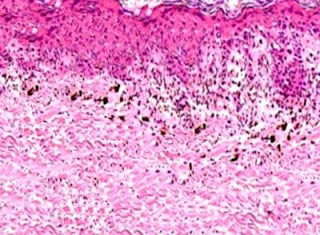
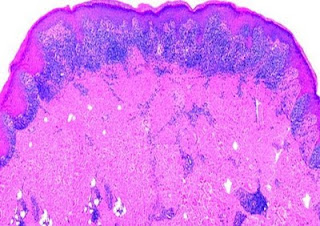
Immunohistochemistry: CD34 is strongly positive. Dermatofibrosarcoma Protuberans Visit: Dermatopathology site Abstract: Cutaneous CD34+ Spindle Cell Neoplasms: Histopathologic Features Distinguish Spindle Cell Lipoma, Solitary Fibrous Tumor, and Dermatofibrosarcoma Protuberans.Am J Dermatopathol. 2010 Jun 17. Spindle cell lipoma (SCL), dermatofibrosarcoma protuberans (DFSP), and solitary fibrous tumors (SFT) are cutaneous CD34+ spindle cell tumors that may exhibit histopathologic and immunophenotypic overlap. We sought ways to reliably distinguish among these lesions even in small or superficial biopsies. Ten morphologic characteristics were analyzed in a group of 5 SCLs, 6 cutaneous SFTs, and 12 DFSPs. SFT and DFSP exhibited extensive histopathologic overlap in small or partial biopsies. However, adnexal entrapment, defined as diffuse proliferation of tumor cells around pilosebaceous and eccrine structures with minimal disruption or expansion of the dermis, was a feature seen in 10 o

Dermatopathology Case 64

Image
Image1 Image2 Image3 Image4 Case 64 A slow growing solitary reddish nodule in a 55 year old male. The nodule is 5 cm in diameter and is located on the right upper arm. Answer

Answer of Dermatopathology Case 63

Cutaneous Sarcoidosis Visit: Dermatopathology site Visit: Pathology of Cutaneous Sarcoidosis Abstract: Sarcoidosis of the skin--a dermatological puzzle: important differential diagnostic aspects and guidelines for clinical and histopathological recognition. J Eur Acad Dermatol Venereol.2010 Feb;24(2):125-37. Epub 2009 Aug 17. Sarcoidosis of the skin may have an extremely heterogeneous clinical presentation, so that the definitions of 'great imitator' and 'clinical chameleon' have long been used. There is, in fact, a large group of skin diseases that can enter the differential diagnosis with cutaneous sarcoid manifestations, either clinically or/and pathologically. As the clinical consequences and the prognosis of these groups of diseases are often very different, it is important to correctly plan the diagnostic workup. The diagnostic process in this case often presents a challenge as no single test is sufficiently specific, so that a certain diagnosis can be only made

Dermatopathology Case 63

Image
Image 1 Image 2 Image3 Image 4 Case 63 A 35 year old male with maculopapular eruption. Patient also has history of acute lymphadenopathy and uveitis. Answer

Answer of Dermatopathology Case 62

Chondroid Syringoma Visit: Dermatopathology site Visit: Pathology of Chondroid Syringoma Abstract: Chondroid syringoma: a rare tumor of the chest wall. Ann Thorac Surg.2010 Mar;89(3):983-5. Chondroid syringoma, an uncommon, slow-growing, benign, sweat-gland tumor located on the upper right chest wall of a 66-year-old woman is presented. This skin adenexal tumor is typically located on the head and neck region. The unusual location of chondroid syringoma made an accurate preoperative diagnosis difficult, and diagnosis was achieved only by excisional biopsy and histopathologic examination. Total surgical excision remains the best therapeutic option to avoid tumor recurrence and close follow-up is recommended because of a rare possibility of malignant transformation and visceral metastases. Chondroid syringoma of the hand.Scand J Plast Reconstr Surg Hand Surg.2009;43(5):291-3. Chondroid syringoma is a rare cutaneous tumour that usually arises in the head and neck region and is rarely see

Dermatopathology Case 62

Image
Image1 Image2 Image 3 Image4 Image4 Case 62 A 40 year old male with a nodule (2.5cm in size) on the forehead. Answer

Dermatopathology Case Index: Case 41 to Case 60

Dermatopathology Quiz - Case Index : Case 41 = Fibrous Papule of the Face (Fibrous Papule of the Nose) Case 42 = Lupus Panniculitis (Lupus Profundus) Case 43 = Subcutaneous Panniculitis-like T-Cell Lymphoma Case 44 = Necrobiotic Xanthogranuloma Case 45 = Lymphomatoid Papulosis Case 46 = Hailey-Hailey Disease Case 47 = Porokeratosis of Mibelli Case 48 = Cutaneous Leiomyoma (Piloleiomyoma) Case 49 = Mycobacterium Marinum Infection (Fish Tank Granuloma ; Swimming Pool Granuloma) Case 50 = Cutaneous Herpes Simplex Infection Case 51 = Prurigo Nodularis Case 52 = Acquired Tufted Angioma Case 53 = Merkel Cell Carcinoma Case 54 = Ichthyosis Vulgaris Case 55 = Discoid Lupus Erythematosus Case 56 = Lichen Planus Case 57 = Erythema Multiforme Case 58 = Fixed Drug Eruption Case 59 = Pigmented Spindle Cell Nevus of Reed. Case 60 = Spitz Nevus

Answer of Dermatopathology Case 61

Targetoid Hemosiderotic Hemangioma (Hobnail Hemangioma) Visit: Dermatopathology site Visit: Targetoid Hemosiderotic Hemangioma (Hobnail Hemangioma) Abstract: Hobnail hemangiomas (targetoid hemosiderotic hemangiomas) are true lymphangiomas.J Cutan Pathol.2004 May;31(5):362-7. BACKGROUND: Hobnail hemangioma (targetoid hemosiderotic hemangioma) is a small benign vascular tumor of the superficial and mid-dermis. In contrast to its well-characterized histology, it has been unclear whether this tumor arises from blood vessel endothelial cells (BECs) or lymphatic vessel endothelial cells (LECs). METHODS: We analyzed 10 hobnail hemangiomas by immunohistochemistry, using the recently described lymphatic endothelial cell marker, D2-40. For comparison, CD31, CD34, and alpha-smooth muscle actin expression were studied in consecutive sections of the paraffin-embedded tissues. RESULTS: In all analyzed vessels, D2-40 labeled exclusively LECs, whereas BECs were consistently negative. In contrast to c

Dermatopathology Case 61

Image
Image1 Image2 Image3 Image4 Case 61 A 45 year old male with a violaceous central papule surrounded by an area of pallor and a brown ring. Answer

Answer of Dermatopathology Case 60

Spitz Nevus Visit: Dermatopathology site Visit: Pathology of Spitz Nevus The spitz nevus: review and update.Clin Plast Surg.2010 Jan;37(1):21-33. The Spitz nevus is a relatively common skin lesion in children and is less commonly seen in adults. The lesion is defined by the presence of distinctive-appearing spindle or epithelioid cells on light microscopy in a recognizable nevus-like pattern. Spitz lesions share features with melanoma on light microscopic examination. When Spitz features are atypical or typical features are absent, distinction from melanoma can be difficult. A spectrum of pathology of Spitz lesions can be found from lesions that are benign and typical to lesions that are atypical with melanoma-like features and frank melanoma. There is significant interobserver variation in interpretation of Spitz lesions. The lack of uniformly applied criteria for distinction of light microscopic grades and the confusion in diagnostic terminology demonstrate the difficulty in the pat

Dermatopathology Case 60

Image
Image1 Image2 Image3 Case 60 A pink coloured papule on the left lower limb of a 19 year old boy. Answer

Answer of Dermatopathology Case 59

Pigmented Spindle Cell Nevus of Reed. Visit: Dermatopathology site Visit: Pigmented Spindle Cell Nevus (Reed Nevus) Abstract Hypopigmented Reed nevus.J Cutan Pathol. 2008 Oct;35 Suppl 1:87-9. Epub 2008 Jun 28. Reed nevus, also named pigmented spindle cell nevus, is a peculiar melanocytic nevus, now regarded as a variant of Spitz nevus by the majority of authors. It is characterized by spindle-shaped melanocytes disposed in nests located in epidermis and papillary dermis. It is usually heavily pigmented, and many melanophages may also be present. Hypopigmented Reed nevus shows all the typical features of conventional pigmented spindle cell nevus, but it does not contain abundant melanin. This variant of Reed nevus is poorly described in literature, so we report five cases of hypopigmented Reed nevus and discuss its clinical and histopathological features. Epithelioid and hyperpigmented melanocytic tumors. An overview. Pathologe. 2007 Nov;28(6):411-21. Spindle cell and epithelioid cell d

Dermatopathology Case 59

Image
Image1 Image2 Image3 Image 4 Case 59 A 20year old female with a well circumscribed deeply pigmented papule on the left thigh. Answer

Answer of Dermatopathology Case 58

Fixed Drug Eruption Visit: Dermatopathology site Abstract: Clinical resemblance of widespread bullous fixed drug eruption to Stevens-Johnson syndrome or toxic epidermal necrolysis: report of two cases.J Formos Med Assoc. 2002 Aug;101(8):572-6. Widespread bullous fixed drug eruption (FDE) is the most severe form of FDE and may be mistaken clinically for Stevens-Johnson syndrome or toxic epidermal necrolysis (SJS/TEN). We report two cases of generalized bullous drug eruption with extensive epidermal necrosis and detachment mimicking SJS/TEN overlap and TEN, respectively. The first patient, a 78-year-old man, developed SJS/TEN-like eruption with widespread dusky red patches and denuded areas shortly after taking multiple nonsteroidal antiinflammatory drugs (NSAIDs). Histopathology showed vacuolar interface dermatitis with numerous necrotic keratinocytes and a superficial and deep perivascular infiltrate containing lymphocytes, eosinophils, neutrophils and melanophages. These findings are

Dermatopathology Case 58

Image
Image1 Image2 Image3 Image4 Case 58 A 42 year old male with oval shaped erythematous lesion on the face. The lesion usually subside leaving a hyperpigmented macule. patient was taking a combination of trimethoprine-sulphamethoxazole. The lesion subsided on withdrawal of the drug and reappeared as soon as the patient started taking the drug. Answer

Answer of Dermatopathology Case 57

Erythema Multiforme Visit: Dermatopathology site Abstract Lichenoid histopathologic changes in patients with clinical diagnoses of exfoliative dermatitis. Am J Dermatopathol.1991 Aug;13(4):358-64. Among 30 patients who received a clinical diagnosis of exfoliative dermatitis and were biopsied between 1982 and 1990, nine showed microscopic features of lichenoid dermatitis. Clinical information was available in eight of these cases. Possible etiologic factors included lymphoma, herpes simplex infection, connective tissue disease, and (in five cases) reactions to drugs. In each instance, microscopic features included a superficial perivascular lymphocytic infiltrate involving the dermal-epidermal interface, vacuolar alteration of the basilar layer, and individually necrotic keratinocytes at all levels of the epidermis. Such microscopic changes are not usually described in connection with exfoliative dermatitis, with the possible exception of those cases related to lichen planus or lupus er

Dermatopathology Case 57

Image
Image1 Image2 Image3 Image4 Image5 Case 57 A 35 year old male with history of respiratory tract infection. Skin lesions occurs on the extensor surfaces of the extremities. The skin lesion is characterized by erythematous macule which expands in size. Later small vesicles develop. Patient had past history of Herpes simplex virus infection. Answer

Answer of Dermatopathology Case 56

Lichen Planus Visit: Dermatopathology site Abstract: Lichen planus and lichenoid reactions of the oral mucosa.Dermatol Ther. 2010 May-Jun;23(3):251-67. Oral lichenoid reactions represent a common end point in response to extrinsic agents (drugs, allergens), altered self-antigens, or superantigens. Oral lichen planus, a common and under-recognized inflammatory disorder, shares many clinical and histopathological features with oral lichenoid drug reaction and oral lichenoid contact reaction. Clinical presentation can vary from asymptomatic white reticular striae to painful erythema and erosions. Cutaneous and additional mucosal involvement is common. Delay in diagnosis of extraoral mucocutaneous lichen planus (LP) results in conjunctival scarring; vaginal stenosis; vulvar destruction; and stricture of the esophagus, urethra, and external auditory meatus. Although the etiology of LP is idiopathic, oral lichenoid reactions may be caused by medications or exogenous agents such as cinnamates

Dermatopathology Case 56

Image
Image1 Image2 Image3 Image4 Image5 Case 56 A 25 year old male with flat pruritic papules on the flexor surface of the wrist. Answer

Answer of Dermatopathology Case 55

Discoid Lupus Erythematosus Visit: Dermatopathology site Abstract: Histologic features of cutaneous lupus erythematosus.Autoimmun Rev.2009 May;8(6):467-73. Epub 2009 Jan 20 Histologic examination of lesions plays a key role in the diagnostics of cutaneous lupus erythematosus (LE). LE has a broad spectrum of histopathological signs, which are related to the stages of the lesions. In addition to the main subtypes of LE, we report on special manifestations like Rowell's-syndrome and Chilblain LE, and give an account of Kikuchi-Fujimoto disease (histiocytic necrotizing lymphadenitis), which may be associated with systemic LE. Furthermore the most considerable histopathologic differential diagnoses are discussed. The different faces of cutaneous lupus erythematosus. G Ital Dermatol Venereol.2009 Apr;144(2):135-47. Lupus erythematosus is a chronic and inflammatory multiorgan disease with variable clinical appearance and variable course. Most patients with systemic lupus erythematosus sho