Posts

Showing posts from 2011

Answer of Dermatopathology Case 111

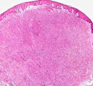
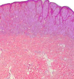
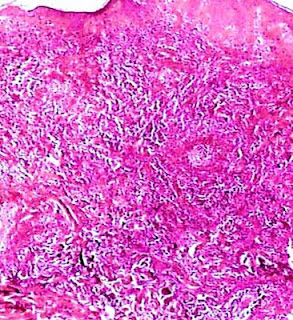
Darier's Disease (Keratosis Follicularis) Visit: Dermatopathology Site Abstract Darier disease: case report with oral manifestations.Med Oral Patol Oral Cir Bucal. 2006 Aug 1;11(5):E404-6. Darier disease, also known as keratosis follicularis or dyskeratosis follicularis, is a rare autosomal dominant genodermatosis. It is clinically manifested by hyperkeratotic papules primarily affecting seborrheic areas on the head, neck and thorax, with less frequent involvement of the oral mucosa. When oral manifestations are present, they primarily affect the palatal and alveolar mucosa, are usually asymptomatic, and are discovered in routine dental examination. Histologically, the lesions present suprabasal clefts in the epithelium with acantholytic and dyskeratotic cells represented by corps ronds and corps grains. This paper reports a case of an adult male patient presenting clinical signs of Darier disease in the palatal mucosa and skin on the neck and upper limbs. Intraoral biopsy of the a

Dermatopathology Case 111

Image
Image1 Image2 Image3 Case 111 A 16 year old boy with yellowish brown,warty papules on the forehead. Small pits are noted on the palms and soles of the feet. Red and white bands are noted in the nails along with V-shaped nick at the free margin of the nail. Diagnosis

Answer of Dermatopathology Case 110

Dermatofibroma with Monster Cells Visit: Dermatopathology Site Visit: Pathology of Dermatofibroma Abstracts: Giant dermatofibroma with monster cells.Am J Dermatopathol. 2002 Feb;24(1):36-8. We report a case of a 64-year-old woman with a giant dermatofibroma on her back with the unusual histologic feature of monster cells. The firm, exophytic, 3-cm nodule had purple and yellow components with surface telangiectasia. Histologic examination demonstrated characteristic findings of a dermatofibroma, including rete ridge flattening and bridging; a stroma containing scattered, large, round, eosinophilic collagen bundles; and a polymorphous dermal infiltrate of spindle and xanthomatous cells with scattered siderophages. Some xanthomatous cells demonstrated features consistent with monster cells, including huge bizarre nuclei and one or more nucleoli. Immunohistochemical staining for factor XIIIa was positive. A diagnosis of giant dermatofibroma with monster cells (DFMC) was made. Giant dermato

Dermatopathology Case 110

Image
Image1 Image2 Image3 Case 110 A 55 year old female with a brownish firm nodule on the back. The nodule is 2.5 cm in diameter. Diagnosis

Answer of Dermatopathology Case 109

Image
HMB-45 : Positive Balloon Cell Melanoma Visit: Dermatopathology Site Visit: Pathology of Balloon Cell Melanoma

Dermatopathology Case 109

Image
Image1 Image2 Image3 Image4 Image5 Case 109 A 72 year old male with a skin lesion on the forehead. Diagnosis

Answer of Dermatopathology Case 108

Median Raphe Cyst Visit: Dermatopathology Site Visit: Pathology of Median Raphe Cyst Abstract: Median raphe cyst of the penis.Dermatol Online J. 2005 Dec 1;11(3):37. Cysts of the median raphe are uncommon. We describe a 43-year-old man with an asymptomatic nodule on the glans penis. Excision of the lesion was performed followed by histological and immunohistochemical studies. Histopathological examination revealed a solitary unilocular cystic cavity in the corium, lined by a pseudostratified columnar epithelium, among which mucinous cells where identified. Hints of decapitation secretion occurred at the apex of the luminal layer. An immunohistochemical study using a standard avidin-biotin peroxidase method disclosed CK7 and CK13 reactivity in the columnar cells. Epithelial membrane antigen (EMA) and carcinoembryonic (CEA) antigen immunoreactivity occurred at the apical border of the luminal cells. No staining was obtained with anti-CK20, human milk fat globulin 1 (HMFG1) and anti- S10

Dermatopathology Case 108

Image
Image1 Image 2 Image3 Image4 Case 108 A 38 year old male with a nodule on the glans penis. Diagnosis

Answer of Dermatopathology Case 107

Naevoid Melanoma Visit: Dermatopathology Site Visit: Pathology of Naevoid Melanoma In general, common naevi are diagnosable at low power. It is important to exclude the possibility of naevoid melanoma. This is a rare variant that mimics benign naevi and is difficult to recognise. The correct diagnosis is frequently made only after the patient has developed a metastasis. At low power, naevoid melanoma may have a nodular architecture, and exhibit other features of a common banal naevus, including circumscription and at least relative symmetry. Naevoid melanoma lacks the prominent junctional activity and pagetoid spread usually associated with superficial spreading melanoma. Common intradermal naevus “matures” (there is an overall decrease in nest size and cellular and nuclear size with depth). On low power examination, naevoid melanoma may appear to mature with depth, yet closer inspection reveals that the cells at the base of the lesions are similar in size to those of the superficial

Dermatopathology Case 107

Image
Image 1 Image 2 Image 3 Image 4 Image 5 Case 107 A 45 year old female with a pigmented lesion on the left side of the chest. Diagnosis

Answer of Dermatopathology Case 106

Desmoplastic Spitz Naevus Visit: Dermatopathology Site Visit: Pathology of Desmoplastic Spitz Naevus Visit: Pathology of Spitz Naevus Abstract: Desmoplastic Spitz nevus: a histopathological review and comparison with desmoplastic melanoma. Ann Dermatol Venereol. 2009 Oct;136(10):689-95. INTRODUCTION: Desmoplastic Spitz nevus is a rare variant of spitzoid tumours characterized by dermal proliferation of large epitheliod and/or fusiform melanocytes within a desmoplastic stroma, comprising thick, eosinophilic collagen bundles. It occurs most frequently in young adults and children, and exhibits a predilection for the limbs. Histologically, this entity may be mistaken for cutaneous fibrous tumours or desmoplastic melanoma. To establish useful histological criteria for differential diagnosis of desmoplastic Spitz nevus, we conducted a retrospective study comparing data concerning the clinical, demographic and histological characteristics of desmoplastic Spitz nevus and desmoplastic melanom

Dermatopathology Case 106

Image
Image1 Image2 Image2 Image3 Image4 Case 106 A 28 year old female with a reddish brown nodule on the left leg. Diagnosis

Answer of Dermatopathology Case 105

Image
Immunostaining, staining reveals that the epithelial cells are positive for cytokeratin , estrogen receptor (ER) and progesterone receptor (PR). PR (Progesterone Receptor):Positive ER (Estrogen Receptor): Positive Cytokeratin: Positive Diagnosis: Cutaneous Ciliated Cyst Visit: Dermatopathology Site Visit: Pathology of Cutaneous Ciliated Cyst Abstract: Cutaneous ciliated cyst of the right lower leg. Pathol Int. 1999 Apr;49(4):354-7. A 23-year-old Japanese woman with a cutaneous ciliated cyst on her right lower leg is reported. A subcutaneous cyst, measuring 2.5 cm in diameter with papillary projections into the lumen, was lined with ciliated cuboidal to columnar epithelia with partial stratification, histologically. These lining cells did not produce mucin. Immunohistochemically, the ciliated lining cells of the cyst were diffusely positive to epithelial membrane antigen and cytokeratin. In addition, positive immunoreaction with anti-desmin monoclonal antibody was observed in the body

Dermatopathology Case 105

Image
Image1 Image2   Case 105 A 38 year old female with a cystic lesion on the lower part of the left leg. The lesion is 2.5cm in diameter. Diagnosis

Answer of Dermatopathology Case 104

Allergic Granulomatosis (Churg-Strauss Syndrome) Visit: Dermatopathology Site Visit: Allergic Granulomatosis (Churg-Strauss Syndrome) Visit: Pathology of Churg-Strauss Syndrome) The three major microscopic features of Allergic Granulomatosis are: 1) Necrotizing vasculitis 2) Tissue infiltration by eosinophils 3) Extravascular granulomas It should be noted that the 3 features mentioned above do not always coexist. In Case No 104 there is small-vessel necrotizing vasculitis together with extensive tissue infiltration by eosinophils. No granulomas were present in the multiple sections taken. Hence in this case detailed clinical history played a very important role to reach the final diagnosis.

Dermatopathology Case 104

Image
Image 1 Image2 Image 3 Image4 Image5 Image6 Case 104 A 42 year old female with history of asthma and allergic rhinitis. There is marked peripheral blood eosinophilia. Erythemous macules are present on the extremities. Biopsy from the skin lesion. Diagnosis

Answer of Dermatopathology Case 103

Infantile Hemangioma Visit: Dermatopathology Site Visit: Pathology of Infantile Hemangioma Abstract: Comparative study on pathology of noninvoluting congenital hemangioma and infantile hemangioma.Zhonghua Zheng Xing Wai Ke Za Zhi. 2011 May;27(3):178-81 OBJECTIVE: To distinguish noninvoluting congenital hemangioma (NICH) and infantile hemangioma (IH) by comparing the pathological structure and marker antigen expression. METHODS: From Jan. 2005 to Aug. 2010, 39 paraffin-embedded samples, including 13 cases of NICH, 13 cases of proliferating IH and 13 cases of involuting IH, were collected from operation. Hematoxylin-eosin staining was used to observe the pathological structure. Immunohistochemical analysis was also performed to investigate the expression of Glut-1. RESULTS: The lobules of capillaries were well-defined in NICH. The lobules were surrounded by abundant fibrous tissue. The capillaries were often large and integrity in NICH. There were few mitosis and apoptosis in endothelial

Dermatopathology Case 103

Image
Image1 Image2 Image3 Image4 Image5 Case 103 A 9 month old child with a reddish nodule on the forehead. Diagnosis

Answer of Dermatopathology Case 102

Acrodermatitis Chronica Atrophicans Visit: Dermatopathology Site Visit: Pathology of Acrodermatitis Chronica Atrophicans There is superficial and deep chronic inflammatory cellular infiltrate in the dermis. It is moderately heavy and composed mainly of lymphocytes together with histiocytes and plasma cells. There is telangiectasia and the cellular infiltrate is prominent around the blood vessels. Inflammatory cells are also present around the adnexae. Thickened collagen bundles are haphazardly present in the upper half of the dermis. There is some atrophy of the dermis and of the subcutis. The epidermis also shows variable atrophy with loss of rete pegs.

Dermatopathology Case 102

Image
Image1 Image2 Image3 Image4 Image5 Case 102 A 68 year old male with ill-defined erythema on the extensor surface of the lower end of the right leg extending to the dorsum of the foot.There is associated wrinkling of the skin. Diagnosis

Dermatopathology Case Index: Case 81 to Case 100

Dermatopathology Quiz - Case Index Case 81 = Langerhans Cell Histiocytosis Case 82 = Rheumatoid Nodule Case 83 = Gouty Tophus Case 84 = Polymorphous Light Eruption Case 85 = Graft-Versus-Host Disease Case 86 = Pilar Sheath Acanthoma Case 87 = Angiokeratoma Case 88 = Neurothekeoma (Nerve Sheath Myxoma) Case 89 = Digital Mucous Cyst Case 90 = Lichen Simplex Chronicus Case 91 = Proliferating Trichilemmal Cyst (Pilar Tumour) Case 92 = Subcorneal Pustular Dermatosis (Sneddon-Wilkinson disease) Case 93 = Urticaria Pigmentosa Case 94 = Hypertrophic Lichen Planus Case 95 = Papillary Eccrine Adenoma Case 96 = Primary Cutaneous Anaplastic Large Cell Lymphoma Case 97 = Mycosis Fungoides-associated Follicular Mucinosis Case 98 = Epithelioid Hemangioendothelioma Case 99 = Kaposiform Hemangioendothelioma Case 100 = Retiform Hemangioendothelioma

Answer of Dermatopathology Case 101

Microvenular Hemangioma Visit: Dermatopathology Site Visit: Pathology of Microvenular Hemangioma Abstract: Microvenular hemangioma. Ann Dermatol Venereol. 2004 May;131(5):475-7. INTRODUCTION: Microvenular hemangioma belongs to the group of acquired vascular tumors. It is important to differentiate such lesions because of their prognostic and nosologic consequences. We report a case of microvenular hemangioma. CASE REPORT: A 31 year-old man presented with a 3 cm erythematous and asymptomatic nodule of the abdomen, which had grown for 2 months. Histopathology showed the irregular dermal proliferation of small vessels, composed of capillaries and venules, without atypia. No relapse was noted 6 months after complete exeresis. DISCUSSION: Microvenular hemangioma is a recently described vascular tumor. The first three cases were reported in 1989, with the denomination of "microcapillar hemangioma". Twenty-one further cases have been reported since 1991. We discuss the typical clin

Dermatopathology Case 101

Image
Image 1 Image2 Image3 Image4 Image5 Case 101 A solitary erythematous nodule on the right forearm of a 45 year old woman. Diagnosis