Posts

Answer of Dermatopathology Case 100

Retiform Hemangioendothelioma Visit: Dermatopathology Site Visit: Pathology of Hemangioendothelioma Abstract: Retiform haemangioendothelioma: a case report.Ann Pathol. 2009;29(6):491-4. Retiform haemangioendothelioma is a locally aggressive, very rarely metastasizing vascular lesion. Histologically, it is characterized by distinctive arborizing blood vessels resembling "rete testis" and lined by endothelial cells with characteristic hobnail morphology. We present an additional case, in the leg of a 64-year-old patient. We discuss the classification of hemangioendotheliomas. The term hemangioendothelioma should be restricted to vascular tumours of "intermediate malignancy" but has been used to designate tumours with variable histological features and clinical behaviour. Spindle cell hemangio(endothelio)ma is currently regarded as a benign reactive lesion. Kaposiform hemangioendothelioma is potentially lethal due to consumption coagulopathy but no metastasizing case h

Dermatopathology Case 100

Image
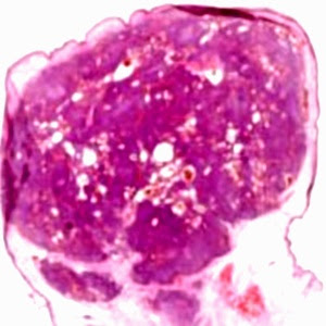
Image 1 Image 2 Image 3 Image4 Case 100 A 28 year old male with an indolent brownish plaque on the right lower limb. Diagnosis

Answer of Dermatopathology Case 99

Kaposiform Hemangioendothelioma Visit: Dermatopathology Site Visit: Pathology of Hemangioendothelioma Abstract: Successful treatment of Kasabach-Merritt syndrome arising from kaposiform hemangioendothelioma by systemic corticosteroid therapy and surgery.Int J Clin Oncol. 2011 Sep 28. Kasabach-Merritt syndrome is a rare type of vascular tumor with aggressive behavior in association with thrombocytopenia and consumptive coagulopathy. A standard guideline has not been established to date. A 7-day-old male infant with Kasabach-Merritt syndrome arising from kaposiform hemangioendothelioma was successfully treated with systemic corticosteroid and surgery. Systemic corticosteroid including methylprednisolone was injected intravenously followed by an intralesional injection of compound betamethasone. This approach brought about an excellent response after the first treatment which was maintained long enough to provide us with an opportunity to excise the tumor. Systemic corticosteroid and surg

Dermatopathology Case 99

Image
Image1 Image2 Image3 Image4 Image5 Case 99 A reddish blue nodule on the left upper arm of a 3 year old child. Diagnosis

Answer of Dermatopathology Case 98

Epithelioid Hemangioendothelioma Visit: Dermatopathology site Visit: Pathology of Hemangioendothelioma Abstract Epithelioid hemangioendothelioma presenting in the skin: a clinicopathologic study of eight cases. Am J Dermatopathol.1998 Dec;20(6):541-6. Epithelioid hemangioendothelioma arising in the skin is extremely rare, and the majority of documented cases have developed in association with an underlying bone tumor. We report eight patients with an age range of 29-84 years (mean 53), who presented with primary cutaneous tumors at a variety of sites including the palm, shin, neck, knee, nose, back, and penis with a duration of between 6 and 12 months. Histologically, all eight cases presented as circumscribed nodules with an overlying acanthotic epidermis, three showing striking acrosyringeal proliferation, reminiscent of eccrine syringofibroadenoma. The tumors were composed of an admixture of slightly pleomorphic spindle and epithelioid cells with abundant, sharply defined eosinophil