Posts

Showing posts from January, 2011

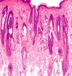
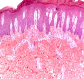
Answer of Dermatopathology Case 85

Image
Graft-Versus-Host Disease Visit: Dermatopathology site Visit: Cutaneous Lesions in Graft versus Host Disease Abstract: Clinicopathologic characteristics of cutaneous chronic graft-versus-host diseases: a retrospective study in Korean patients. Int J Dermatol. 2010 Dec;49(12):1386-92. BACKGROUND: Chronic graft-versus-host disease (cGVHD) is a major complication in long-term survivors of hematopoietic stem cell transplantation (HSCT). Cutaneous manifestations are frequently the presenting features; therefore, the dermatologist needs to be aware of the wide spectrum of cutaneous cGVHD. METHODS: We retrospectively evaluated patients' characteristics, clinical, and histological features of cutaneous cGVHD and analyzed factors influencing the severity of cutaneous cGVHD in 100 Korean HSCT recipients between January 1, 1995, and December 31, 2007. RESULTS: Clinical manifestations of cutaneous cGVHD mainly presented as lichenoid (60.0%), sclerodermoid (12.0%), or erythematous maculopapul

Dermatopathology Case 85

Image
Image1 Image2 Image3 Case 85 A 35 year old male with skin lesions. Patient has a history of hematopoietic stem cell transplantation. Diagnosis

Answer of Dermatopathology Case 84

Polymorphous Light Eruption Visit: Dermatopathology site Abstracts Polymorphous light eruption.Photodermatol Photoimmunol Photomed.2008 Jun;24(3):155-61. Polymorphous light eruption is the most common photodermatosis, with a prevalence of as high as 10-20% in Western Europe and in the USA. It starts during the second and third decades of life. Although not life-threatening it can severely impair the quality of life, in particular during leisure activities and in outdoors workers. Polymorphous light eruption belongs to the group of so-called idiopathic photodermatoses. This term denotes dermatoses that occur in otherwise healthy individuals from exposure to sunlight or artificial light without the intervention of an exogenous photosensitizing agent. These diseases have two factors in common: they are precipitated by ultraviolet or visible radiation; and their exact pathomechanism remains obscure but is presumably immunologic in nature. Marked papillary dermal edema--an unreliable discr

Dermatopathology Case 84

Image
Image1 Image2 Case 84 Small pruritic papules on the dorsum of the hands and forearms of a 25 year old male. The patient has a history of exposure to bright sunlight for many hours. Answer

Answer of Dermatopathology Case 83

Gouty Tophus Visit: Dermatopathology site Asymptomatic nodule on an elderly lady's thumb tip. Hautarzt. 2011 Jan 26. A 71-year-old woman presented with an asymptomatic growing dermal tumor on her thumb. Clinical picture, ultrasound, laboratory investigations and histology were consistent with the diagnosis of gouty tophus. Pathogenesis, risk factors and therapy of tophaceous gout are discussed. Gouty tophus of the upper eyelid. Ophthal Plast Reconstr Surg. 2008 Sep-Oct;24(5):404-6. A 64-year-old man with gout presented with history of an enlarging mass in the left upper eyelid causing mechanical ptosis. The patient underwent excisional biopsy. The histopathologic findings confirmed the diagnosis of gouty tophus of the eyelid. Role of Melanocortin Receptors in the Regulation of Gouty Inflammation. Curr Rheumatol Rep. 2011 Jan 18. Gouty arthritis is a form of acute joint inflammation provoked by joint deposition of urate crystals. Although this acute pathology resolves after a few da

Dermatopathology Case 83

Image
Image1 Image2 Image3 Case 83 Slide for spot diagnosis. Answer

Answer of Dermatopathology Case 82

Rheumatoid Nodule Visit: Dermatopathology site Non-infectious granulomatous diseases of the skin and their associated systemic diseases: an evidence-based update to important clinical questions. Am J Clin Dermatol. 2010;11(3):171-81. Non-infectious granulomatous diseases of the skin are a broad group of distinct reactive inflammatory conditions that share important similarities. As a group, they are relatively difficult to diagnose and distinguish both clinically as well as histologically. Many of these disorders have significant associations with systemic diseases that impact the patient's overall prognosis. In this update, we offer a discussion of emerging concepts and controversies in this field, as presented through evidence-based answers to seven important clinical questions regarding palisading and epithelioid granulomata. These questions offer an opportunity to review ten non-infectious granulomatous conditions that have implications for systemic disease: granuloma annulare,

Dermatopathology Case 82

Image
Image1 Image2 Image3 Image4 Case 82 Slide for spot diagnosis. Answer

Answer of Dermatopathology Case 81

Image
Immunohistochemistry: S100 protein is strongly positive. Immunohistochemistry: CD1a is stongly positive Langerhans Cell Histiocytosis Visit: Dermatopathology site Abstract: Langerhans cell hyperplasia of the skin mimicking Langerhans cell histiocytosis: a report of two cases in children not associated with scabies. Fetal Pediatr Pathol. 2010;29(4):231-8. Langerhans cells histiocytosis (LCH) affecting the skin most commonly has clinical and histopathologic diagnostic features. We are reporting two examples of Langerhans cell (LC) hyperplasia recognized in the skin biopsies of two children initially interpreted as LCH. The first was an 8-year-old boy finally interpreted as having an atypical type of contact dermatitis, while the second, an 8-year-old girl, was assumed to have Pytiriasis lichenoides et varioliformis acuta. None showed evidences of scabies. Both presented spongiotic dermatitis with numerous CD1a+ cells. As more cases of LC hyperplasia are recognized, new details emerge h

Dermatopathology Case 81

Image
Image1 Image2 Image3 Image4 Case 81 A 4 year old child with multiple yellow-brown scaly papules on the scalp. Erythematous macules and papules are also present on the neck and back. The child is small for age and there is marked loss of weight. He is also accompanied by anaemia, fever and lymphadenopathy. Answer

Dermatopathology Case Index: Case 61 to Case 80

Dermatopathology Quiz - Case Index Case 61 = Targetoid Hemosiderotic Hemangioma (Hobnail Hemangioma) Case 62 = Chondroid Syringoma Case 63 = Cutaneous Sarcoidosis Case 64 = Dermatofibrosarcoma Protuberans Case 65 = Trichilemmal Carcinoma Case 66 = Melanocytic Nevus of the Vulva (Site specific nevus) Case 67 = Lichen Amyloidosus Case 68 = Scabies Case 69 = Dermatophyte Infection (Dermatophytoses) - Superficial filamentous Infection (Tinea - ringworm) Case 70 = Cutaneous Malakoplakia Case 71 = Grover's Disease Case 72 = Cutaneous Leishmaniasis Case 73 = Cutaneous Histoplasmosis Case 74 = Atrophie Blanche (Livedoid Vasculopathy) Case 75 = Granuloma Faciale Case 76 = Perniosis (Chilblains) Case 77 = Psoriasis Case 78 = Pustular Psoriasis Case 79 = Alopecia Areata Case 80 = Nodular Vasculitis (Erythema Induratum, Bazin-type)

Answer of Dermatopathology Case 80

Nodular Vasculitis (Erythema Induratum, Bazin-type) Visit: Dermatopathology site Abstract: Erythema induratum of Bazin. Dermatol Online J.2010 Apr 15;16(4):1. An 81-year-old woman with a history of renal cell carcinoma and years of slowly, progressively enlarging pulmonary nodules of uncertain etiology presented with several weeks of painful lower extremity nodules. A biopsy revealed changes consistent with nodular vasculitis. A purified protein derivative and QuantiFERON test were positive, favoring the diagnosis of erythema induratum of Bazin. Treatment with a standard four-drug antituberculous regimen resulted in radiographic and clinical improvement. This case emphasizes the importance of dermatologic manifestations in the detection of systemic disease. Vasculitis in erythema induratum of Bazin: a histopathologic study of 101 biopsy specimens from 86 patients. J Am Acad Dermatol. 2008 Nov;59(5):839-51. BACKGROUND: Erythema induratum of Bazin is a mostly lobular panniculitis. There

Dermatopathology Case 80

Image
Image1 Image2 Image 3 Image4 Case 80 A 55 year old woman with firm, tender erythematous nodules on the calves. Answer

Answer of Dermatopathology Case 79

Alopecia Areata Visit: Dermatopathology site Abstract: 'Follicular Swiss cheese' pattern--another histopathologic clue to alopecia areata.J Cutan Pathol. 2011 Feb;38(2):185-9. Yellow dots are the most useful dermoscopic criterion in the clinical diagnosis of alopecia areata and correspond histopathologically with dilated follicular infundibula. They are found in about 95% of alopecia areata cases and help to differentiate alopecia areata from trichotillomania, telogen effluvium and from scarring alopecias. Histopathology of alopecia areata differs with disease activity and dermatopathologist, therefore, heavily depends on other diagnostic features. Objective of the study was to determine the frequency of dilated follicular infundibula, peribulbar lymphocytic infiltrate, inflammatory infiltrates of lymphocytes and eosinophils within fibrous streamers and a shift to catagen/telogen follicles in alopecia areata. Histopathologic features of 56 specimens of 33 patients were correlat

Dermatopathology Case 79

Image
Image1 Image 2 Image3 Image4 Case 79 A 24 year old male with a few small round patches of hair loss on the scalp. "Exclamation-mark" hairs are seen at the margin of the patch. Sections of the scalp biopsy from the patch. Answer

Answer of Dermatopathology Case 78

Pustular Psoriasis Visit: Dermatopathology site Abstracts: The histopathological spectrum of acute generalized exanthematous pustulosis (AGEP) and its differentiation from generalized pustular psoriasis. J Cutan Pathol. 2010 Dec;37(12):1220-9. BACKGROUND: Acute generalized exanthematous pustulosis (AGEP) represents a severe, acute, pustular skin reaction that is most often induced by drugs. AGEP can be difficult to differentiate from generalized pustular psoriasis (GPP) both clinically and histopathologically. We present a systematic description of the histopathological spectrum of AGEP and GPP with a focus on discriminating features. MATERIALS AND METHODS: A retrospective, descriptive, comparative histopathological study was completed utilizing step sections of 43 biopsies of 29 cases with a validated diagnosis of probable or definite AGEP and 24 biopsies of 19 cases with an established diagnosis of GPP. RESULTS: In AGEP, biopsies from erythema and pustules showed minor differences, w

Dermatopathology Case 78

Image
Image1 Image2 Image 3 Case 78 A 50 year old male with clearly defined pustules on the skin. The lesions are painful. The patient also complains of fever and malaise. Answer

Answer of Dermatopathology Case 77

Psoriasis Visit: Dermatopathology site Abstract: Scalp psoriasis: an overview of the disease and available therapies.J Drugs Dermatol. 2010 Aug;9(8):912-8. The scalp is one of the regions of the body most commonly affected by psoriatic lesions. While the head represents only 10 percent of the body's surface area, the consequences of scalp psoriasis are disproportionate to the area, as it can be seriously debilitating and presents social and emotional distress to the affected individual. Scalp lesions are often well-demarcated and may have thick gray or white scale; patients with scalp psoriasis frequently complain of pruritus and shedding of scale. Current treatment modalities--including phototherapy, topical corticosteroids, topical vitamin D analogues and conventional systemic therapies--have produced unsatisfactory results for patients with moderate-to-severe scalp psoriasis due to difficulties in administration to the disease site, poor compliance, toxicity and inadequate long-

Dermatopathology Case 77

Image
Image 1 Image2 Image 3 Case 77 Small red dry patches on both elbows of a 45 year old woman. Patient complains of intense itching. Answer

Answer of Dermatopathology Case 76

Perniosis (Chilblains) Visit: Dermatopathology site Abstract: Perniosis: clinical and histopathological analysis. Am J Dermatopathol. 2010 Feb;32(1):19-23. Perniosis are inflammatory cutaneous lesions, located on acral skin, which present in association with cold exposure. They can appear as an idiopathic dermatosis or with an underlying autoimmune disease. The use of cutaneous biopsy to distinguish between both types is controversial. We analyze the histological findings in 9 cases of idiopathic perniosis (IP) and compare them with those obtained from 11 cases of perniosis associated with an autoimmune disease (autoimmune perniosis). The most frequent histopathological features observed in cases of IP were a lymphocytic infiltrate with perivascular (8 cases, 89%) and perieccrine distribution (6 cases, 66%), dermal edema (5 cases, 55%), and necrotic keratinocytes (5 cases, 55%), whereas those found in perniosis associated with an autoimmune disease were lymphocytic infiltrate with peri

Dermatopathology Case 76

Image
Image1 Image2 Image3 Image4 Case 76 A 19 year old girl with bluish red, painful and itchy papules on the toes. She was exposed to extremely cold temperature in the winter season. Answer

Answer of Dermatopathology Case 75

Granuloma Faciale Visit: Dermatopathology site Abstract: Granuloma faciale with disseminated extra facial lesions. Dermatol Online J.2010 Jun 15;16(6):5. Granuloma faciale (GF) is a rare cutaneous disorder categorized as a localized form of small vessel vasculitis. Clinically, it manifests as single or multiple, well-demarcated, red-brown plaques, papules and nodules, nearly always confined to the face. Herein, we report a 39-year-old man with multiple red-brown, infiltrated plaques on his face and extrafacial lesions on the back, shoulders, and both arms. Skin biopsy revealed typical histopathological findings of GF. The patient failed to respond to pulsed dye laser, but intralesional triamcinolone combined with cryotherapy led to an acceptable response. Granuloma faciale: Case report and review. Dermatol Online J. 2009 Dec 15;15(12):3. Granuloma faciale (GF) is a rare benign chronic inflammatory dermatosis usually appearing only on the face. The lesions of GF typically present as sin