Posts

Answer of Dermatopathology Case 110

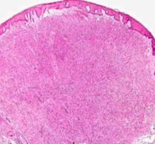
Dermatofibroma with Monster Cells Visit: Dermatopathology Site Visit: Pathology of Dermatofibroma Abstracts: Giant dermatofibroma with monster cells.Am J Dermatopathol. 2002 Feb;24(1):36-8. We report a case of a 64-year-old woman with a giant dermatofibroma on her back with the unusual histologic feature of monster cells. The firm, exophytic, 3-cm nodule had purple and yellow components with surface telangiectasia. Histologic examination demonstrated characteristic findings of a dermatofibroma, including rete ridge flattening and bridging; a stroma containing scattered, large, round, eosinophilic collagen bundles; and a polymorphous dermal infiltrate of spindle and xanthomatous cells with scattered siderophages. Some xanthomatous cells demonstrated features consistent with monster cells, including huge bizarre nuclei and one or more nucleoli. Immunohistochemical staining for factor XIIIa was positive. A diagnosis of giant dermatofibroma with monster cells (DFMC) was made. Giant dermato

Dermatopathology Case 110

Image
Image1 Image2 Image3 Case 110 A 55 year old female with a brownish firm nodule on the back. The nodule is 2.5 cm in diameter. Diagnosis

Answer of Dermatopathology Case 109

Image
HMB-45 : Positive Balloon Cell Melanoma Visit: Dermatopathology Site Visit: Pathology of Balloon Cell Melanoma

Dermatopathology Case 109

Image
Image1 Image2 Image3 Image4 Image5 Case 109 A 72 year old male with a skin lesion on the forehead. Diagnosis

Answer of Dermatopathology Case 108

Median Raphe Cyst Visit: Dermatopathology Site Visit: Pathology of Median Raphe Cyst Abstract: Median raphe cyst of the penis.Dermatol Online J. 2005 Dec 1;11(3):37. Cysts of the median raphe are uncommon. We describe a 43-year-old man with an asymptomatic nodule on the glans penis. Excision of the lesion was performed followed by histological and immunohistochemical studies. Histopathological examination revealed a solitary unilocular cystic cavity in the corium, lined by a pseudostratified columnar epithelium, among which mucinous cells where identified. Hints of decapitation secretion occurred at the apex of the luminal layer. An immunohistochemical study using a standard avidin-biotin peroxidase method disclosed CK7 and CK13 reactivity in the columnar cells. Epithelial membrane antigen (EMA) and carcinoembryonic (CEA) antigen immunoreactivity occurred at the apical border of the luminal cells. No staining was obtained with anti-CK20, human milk fat globulin 1 (HMFG1) and anti- S10