Posts

Dermatopathology Case 95

Image
Image1 Image2 Image3 Image4 Image5 Case 95 A 45 year old male with a firm nodule on the dorsum of his right hand. Diagnosis

Answer of Dermatopathology Case 94

Image
Hypertrophic Lichen Planus Visit: Dermatopathology site [ Related links to clinical images of similar condition: (DermAtlas -JHU.): Image1 ; Image2 ; Image3 ; Image4 ] Abstract: Verrucous squamous cell carcinoma complicating hypertrophic lichen planus : Three case reports and review of the literature. Hautarzt. 2011 Jan;62(1):40-45. Lichen planus is a chronic mucocutaneous T-cell-mediated disease, whose cause is still unknown. The first case of lichen planus that transformed into squamous cell carcinoma was reported in 1903. We present three patients in whom squamous cell carcinomas were identified in chronic lichen planus. The world literature includes at least 91 cases, including our three cases. In an epidemiological study, no significant risk of transformation of cutaneous lichen planus into squamous cell carcinomas was found. In contrast, there is a significantly higher risk of malignant transformation in mucosal lichen planus, so that the WHO had graded mucosal lichen planus a

Dermatopathology Case 94

Image
Image1 Image2 Image3 Image4 Case 94 A 20 year old male with multiple pruritic plaques on the extremities. The lesions have a verrucous appearance. Diagnosis [ Related links to clinical images of similar condition: (DermAtlas -JHU.): Image1 ; Image2 ; Image3 ; Image4 ]

Answer of Dermatopathology Case 93

Image
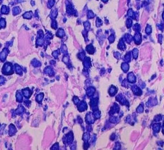
Giemsa stain Toluidine Blue stain Urticaria Pigmentosa Visit: Dermatopathology site Related links to clinical images of the condition: (DermAtlas -JHU.): Image1 ; Image2 ; Image3 ; Image4 . Abstract: Diagnostic criteria and classification of mastocytosis: a consensus proposal. Leuk Res. 2001 Jul;25(7):603-25. The term 'mastocytosis' denotes a heterogeneous group of disorders characterized by abnormal growth and accumulation of mast cells (MC) in one or more organ systems. Over the last 20 years, there has been an evolution in accepted classification systems for this disease. In light of such developments and novel useful markers, it seems appropriate now to re-evaluate and update the classification of mastocytosis. Here, we propose criteria to delineate categories of mastocytosis together with an updated consensus classification system. In this proposal, the diagnosis cutaneous mastocytosis (CM) is based on typical clinical and histological skin lesions and absence of def

Dermatopathology Case 93

Image
Image1 Image2 Image3 Image4 Image5 Case 93 A 2 year old child with multiple reddish brown macules on the trunk. Diagnosis and special stains : [ Related links to clinical images of similar condition: (DermAtlas -JHU.): Image1 ; Image2 ; Image3 ; Image4 .]