Posts

Answer of Dermatopathology Case 111

Darier's Disease (Keratosis Follicularis) Visit: Dermatopathology Site Abstract Darier disease: case report with oral manifestations.Med Oral Patol Oral Cir Bucal. 2006 Aug 1;11(5):E404-6. Darier disease, also known as keratosis follicularis or dyskeratosis follicularis, is a rare autosomal dominant genodermatosis. It is clinically manifested by hyperkeratotic papules primarily affecting seborrheic areas on the head, neck and thorax, with less frequent involvement of the oral mucosa. When oral manifestations are present, they primarily affect the palatal and alveolar mucosa, are usually asymptomatic, and are discovered in routine dental examination. Histologically, the lesions present suprabasal clefts in the epithelium with acantholytic and dyskeratotic cells represented by corps ronds and corps grains. This paper reports a case of an adult male patient presenting clinical signs of Darier disease in the palatal mucosa and skin on the neck and upper limbs. Intraoral biopsy of the a...

Dermatopathology Case 111

Image
Image1 Image2 Image3 Case 111 A 16 year old boy with yellowish brown,warty papules on the forehead. Small pits are noted on the palms and soles of the feet. Red and white bands are noted in the nails along with V-shaped nick at the free margin of the nail. Diagnosis

Answer of Dermatopathology Case 110

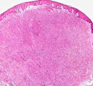
Dermatofibroma with Monster Cells Visit: Dermatopathology Site Visit: Pathology of Dermatofibroma Abstracts: Giant dermatofibroma with monster cells.Am J Dermatopathol. 2002 Feb;24(1):36-8. We report a case of a 64-year-old woman with a giant dermatofibroma on her back with the unusual histologic feature of monster cells. The firm, exophytic, 3-cm nodule had purple and yellow components with surface telangiectasia. Histologic examination demonstrated characteristic findings of a dermatofibroma, including rete ridge flattening and bridging; a stroma containing scattered, large, round, eosinophilic collagen bundles; and a polymorphous dermal infiltrate of spindle and xanthomatous cells with scattered siderophages. Some xanthomatous cells demonstrated features consistent with monster cells, including huge bizarre nuclei and one or more nucleoli. Immunohistochemical staining for factor XIIIa was positive. A diagnosis of giant dermatofibroma with monster cells (DFMC) was made. Giant dermato...

Dermatopathology Case 110

Image
Image1 Image2 Image3 Case 110 A 55 year old female with a brownish firm nodule on the back. The nodule is 2.5 cm in diameter. Diagnosis

Answer of Dermatopathology Case 109

Image
HMB-45 : Positive Balloon Cell Melanoma Visit: Dermatopathology Site Visit: Pathology of Balloon Cell Melanoma