Posts

Dermatopathology Case 130

Image
 Image1  Image2  Image3  Image4 Case 130   A reddish gray nodule, 2cm in diameter, on the forehead of a 20 year old male. Diagnosis

Dermatopathology Case 129

Image
 Image1 Image2  Image3  Image4 Case 129    A nodule on the scalp of a 25 year old male. Diagnosis

Dermatopathology Case 128

Image
Image 1  Image2  Image3  Image4 Case 128 : Annular papule on the right side of the neck of a 45 year old male. Diagnosis

Dermatopathology Case 127

Image
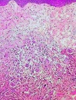
Image 1  Image 2  Image 3 Case 127:   A 21 year old male with history of swollen lips and chronic inflammatory bowel disease. Painful erythematous nodule noted on the left lower leg.    Diagnosis

Dermatopathology Case 126

Image
Image1 Image2 Image3 Case 126:  A 35 year old male with yellowish papule on the eyelid. Diagnosis

Dermatopathology Case 125

Image
Image1 Image2 Image3   Image4 Case 125 -  A 7 year old boy with a pustular lesion on the nose. He has a history of nasal discharge. Diagnosis

Answer of Dermatopathology Case 124

Diagnosis: Pancreatic Panniculitis Visit: Pancreatic Panniculitis

Dermatopathology Case 124

Image
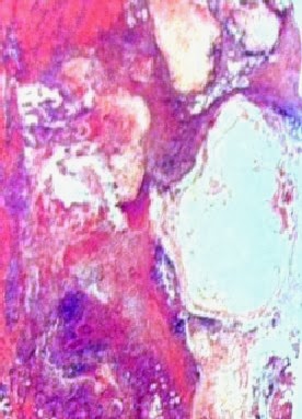
Image1 (Low power)                                             Image2  (Low power)             Image3 (High power)         Image4 (High power)   Case 124: A 45 year old man with painful reddish brown nodule on the left thigh. Patient also has a history of acute pancreatitis.    Spot Diagnosis:

Answer of Dermatopathology Case 123

Answer of Dermatopathology Case 123 : Leukocytoclastic Vasculitis   Visit: Vasculopathic Reaction Pattern

Dermatopathology Case 123

Image
Image1 Image2 Image3 Image4 Case 123   A 35 year old male with an erythematous macule on the left lower leg. Answer

Answer of Dermatopathology Case 122

Diagnosis: Nummular Dermatitis Visit: Spongiotic Reaction Pattern

Dermatopathology Case 122

Image
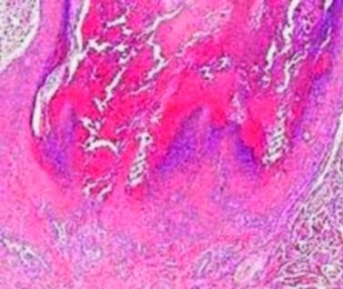
Image1   Image2 Image3   Image 4 Case 122 Coin shaped multiple patches. The surface is crusted. The lesion is located on the left upper arm of a 50 year old male. Answer

Answer of Dermatopathology Case 121

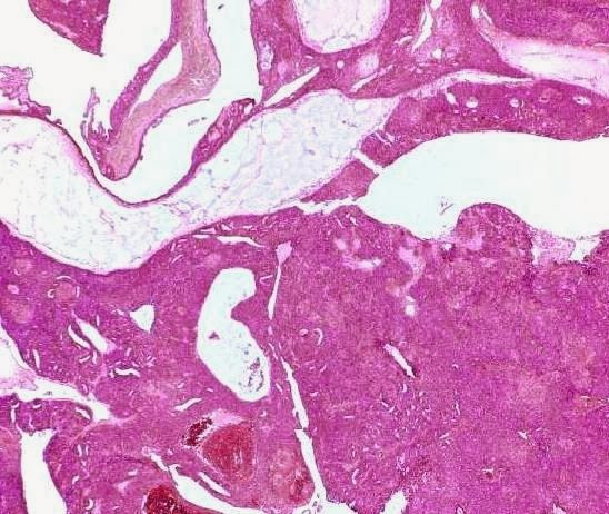
Answer:  Aggressive digital papillary adenocarcinoma (Digital  Papillary Adenocarcinoma)  For further information: Visit: Aggressive digital papillary adenocarcinoma

Dermatopathology Case 121

Image
Image1 Image2 Image3 Image4 Image5 Image6 Case 121:   A  68 year old male with a tan-gray, solitary cystic nodule, 1.5cm in diameter on the left middle finger. Answer:  

Answer of Dermatopathology Case 120

Answer:   Warty Dyskeratoma For further information:  Visit-  Warty Dyskeratoma

Dermatopathology Case 120

Image
Image1 Image2 Image3 Image4 Image5 Case 120 -  A 50 year old man with a solitary nodule on the forehead. Answer:

Answer of Dermatopathology Case 119

Answer:  Bowen's Disease For further information:  Visit:   Bowen's Disease

Dermatopathology Case 119

Image
Image1 Image2 Image3 Image4 Case 119   A 65 year old male, with a red, scaly patch on the left upper arm. Answer:

Answer of Dermatopathology Case 118

Answer: Halo Naevus ( Sutton Nevus) For more information visit: Halo Naevus (Nevus with halo ; Sutton Nevus)

Dermatopathology Case 118

Image
Image1 Image2 Image3 Image4 Case 118  A reddish brown papule on the back of a 14 year old boy. The lesion is surrounded by a white depigmented ring. Answer :