Posts

Dermatopathology Case 53

Image
Image 1 Image2 Image3 Image4 Case 53 A 74 yeare old woman with a nodule on the head. Answer

Answer of Dermatopathology Case 52

Acquired Tufted Angioma Visit: Dermatopathology site Visit: Pathology of Acquired Tufted Angioma Abstract: Perianal acquired tufted angioma associated with pregnancy: case report. Tech Coloproctol. 2002 Sep;6(2):117-9. Tufted angiomas are rare lesions described as slowly growing/spreading erythematous macules especially located in the upper trunk and neck. Herein we report the case of perianal location of a tufted angioma in a young pregnant woman. She came to our observation complaining of perianal pain accompanied by bleeding at defecation. A lesion resembling a perianal fissure was observed. Mild hypertonia of the internal sphincter was confirmed at manometry. After one week of ineffective medical treatment, surgery was planed at the end of the sixteenth week under local anaesthesia. The lesion was excised and a minimal sphincterotomy was performed; histopathology report described features of a tufted angioma. The pregnancy proceeded regularly, without anal symptoms, followed by nor

Dermatopathology Case 52

Image
Image1 Image2 Image3 Case 52 A 10 year old boy with a raised papule on the neck. ? Pyogenic granuloma. Answer

Answer of Dermatopathology Case 51

Prurigo Nodularis Visit: Dermatopathology site Visit: Pathology of Prurigo Nodularis Abstract: Prurigo nodularis: systematic analysis of 58 histological criteria in 136 patients.J Cutan Pathol. 2010 May;37(5):578-86. Epub 2009 Nov 30. BACKGROUND: To date, there has been no systematic investigation of the detailed histological features of prurigo nodularis (PN) in a large cohort of patients. METHODS: This retrospective study includes skin biopsies of 136 patients (63 males, 73 females; mean age: 58.38 years) with PN. RESULTS: Highly characteristic for PN is the presence of thick compact orthohyperkeratosis; the hairy palm sign (folliculosebaceous units in nonvolar skin in conjunction with a thick and compact cornified layer, like that of volar skin); irregular epidermal hyperplasia or pseudoepitheliomatous hyperplasia; focal parakeratosis; hypergranulosis; fibrosis of the papillary dermis with vertically arranged collagen fibers; increased number of fibroblasts and capillaries; a superf

Dermatopathology Case 51

Image
Image1 Image2 Case 51 A 35 year old male, with a few intensely pruritic, firm pink nodules on the extensor aspect of the left upper arm. Answer

Answer of Dermatopathology Case 50

Cutaneous Herpes Simplex Infection Visit: Dermatopathology site Visit: Herpes Simplex Infection Abstract: Histopathology of the more common viral skin infections. Actas Dermosifiliogr .2010 Apr;101(3):201-16. We describe the histopathological characteristics of viral skin infections. Herpes simplex virus and varicella-zoster virus produce an intraepidermal vesicle with variable degrees of epithelial necrosis. Typical findings include keratinocytes with ballooned nuclei with a ground-glass appearance and giant multinucleated keratinocytes. In the endothelial cells of the dermal blood vessels, cytomegalovirus produces large eosinophilic nuclear inclusions surrounded by a clear halo. Human herpes virus 8 is etiologically associated with Kaposi sarcoma. In its early stages, this tumor contains blood vessels with a fine endothelium passing through the dermal collagen bundles. In the plaque and nodular stages, the vessel lumens are more clearly visible and there is a progressive increase in

Dermatopathology Case 50

Image
Image1 Image2 Case 50 A 30 year old immunocompromised male patient with a few painful clear vesicles on the chin. Answer

Answer of Dermatopathology Case 49

Image
Ziehl-Neelsen staining revealed acid-fast bacilli Mycobacterium Marinum Infection (Fish Tank Granuloma ; Swimming Pool Granuloma) Abstract: Fish-Tank Granuloma N Engl J Med 1997; 336:1065April 10, 1997 Fish tank granuloma. BMJ. 1990 April 21; 300(6731): 1069–1070. Fish tank granuloma: misdiagnosed as cutaneous leishmaniasis. Int J Dermatol. 2010 Jan;49(1):53-5. Mycobacterium marinum is an atypical mycobacterium that causes a skin infection known as fish tank granuloma or swimming pool granuloma affecting people who are exposed to aquatic environments. In general, it is managed medically with antimicrobials and variable treatment protocols. Here, we report a Saudi gentleman who acquired this infection in Thailand and was misdiagnosed as cutaneous leishmaniasis. After establishing the correct diagnosis, treatment with minocycline and trimethoprim-sulfamethoxazole resulted in rapid healing. Mycobacterium marinum infections in transplant recipients: case report and review of the literatur

Dermatopathology Case 49

Image
Image1 Image2 Image3 Case 49 A 45 year old fisherman with verrucoid nodules on the elbow and forearm. Answer

Answer of Dermatopathology Case 48

Image
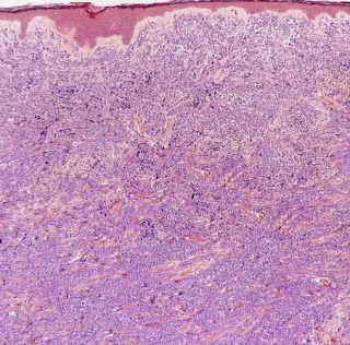
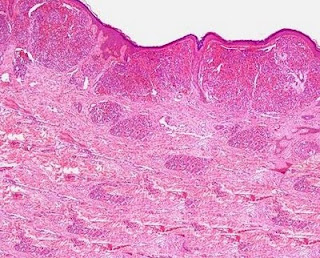
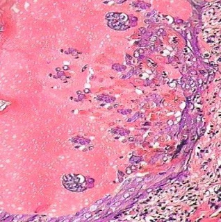
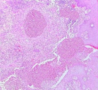
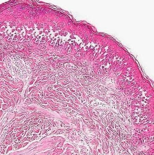
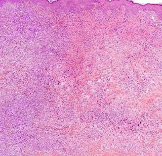
Immunohistochemistry: Smooth Muscle Actin Cutaneous Leiomyoma (Piloleiomyoma) Visit: Dermatopathology site Visit: Pathology of Piloleiomyoma Abstract: Cutaneous smooth muscle neoplasms: clinical features, histologic findings, and treatment options.J Am Acad Dermatol. 2002 Apr;46(4):477-90; quiz, 491-4. Cutaneous smooth muscle is present in 3 separate locations: arrector pili muscles, blood vessel walls, and genital/areolar skin. Benign or malignant smooth muscle neoplasms may arise from each of these locations. This review discusses the pathogenesis, clinical manifestations, histologic findings, prognosis, treatment options, and controversial areas of cutaneous smooth muscle neoplasms. ( J Am Acad Dermatol 2002;46:477-90.) Learning objective: At the completion of this learning activity, participants should be able to discuss the pathogenesis, clinical manifestations, histologic findings, prognosis, and treatment options of cutaneous smooth muscle neoplasms. Case for diagnosis: (unilat

Dermatopathology Case 48

Image
Image1 Image2 Image3 Image4 Case48 A 35 year old male with multiple painful , firm, reddish-brown papulonodules on the extensor surfaces of the extremities. Answer

Answer of Dermatopathology Case 47

Porokeratosis of Mibelli Visit: Dermatopathology site Abstract: Mibelli revisited: a case of type 2 segmental porokeratosis from 1893. J Am Acad Dermatol.2010 Jan;62(1):136-8. Epub 2009 Jul 25. In autosomal dominant skin disorders, a pronounced mosaic involvement may sometimes be found to be superimposed on the ordinary nonsegmental lesions. Such "type 2 segmental manifestation" reflects loss of heterozygosity occurring at an early developmental stage, giving rise to a cell clone that lacks the corresponding wild-type allele. Here, this genetic concept is applied to an unusual case of plaque-type porokeratosis of Mibelli (PM) as published in 1893 by Vittorio Mibelli in the International Atlas of Rare Skin Diseases. The right forearm and hand of the 21-year-old patient showed a pronounced linear porokeratosis that had developed since the age of 2 years. Moreover, nonsegmental lesions of PM involved both hands and forearms as well as the face and the neck, having first been not

Dermatopathology Case 47

Image
Image 1 Image2 Image3 Case 47 A 7 year old girl, with a skin lesion (oval plaque with an atropic center and a thin elevated keratotic rim), on the left forearm. Answer

Answer of Dermatopathology Case 46

Hailey-Hailey Disease Visit: Dermatopathology site Visit: Pathology of Hailey-Hailey Disease Abstract: Familial benign chronic pemphigus (Hailey-Hailey disease) Dermatol Online J. 2009 Aug 15;15(8):15. A case of eczema herpeticum with hailey-hailey disease. Ann Dermatol. 2009 Aug;21(3):311-4. Reevaluation of the normal epidermal calcium gradient, and analysis of calcium levels and ATP receptors in Hailey-Hailey and Darier epidermis. J Invest Dermatol. 2009 Jun;129(6):1379-87. Epub 2008 Dec 4. Electron probe microanalysis was used to analyze elemental content of human epidermis. The results revealed that the calcium content of the basal keratinocyte layer was higher than that of the lowest spinous cell layer in normal epidermis. This was surprising, as it is generally accepted that the calcium level increases with cellular differentiation from the proliferative basal layer to the stratum corneum. Hailey-Hailey disease (HHD) and Darier disease (DD) are caused by mutations in Ca(2+)-ATPa

Dermatopathology Case 46

Image
Image1 Image2 Image3 Case 46 A well demarcated bullous lesion in the axilla of a 48 year old female. Answer

Answer of Dermatopathology Case 45

Lymphomatoid Papulosis Visit: Dermatopathology site Visit: Pathology of Lymphomatoid Papulosis Abstracts: Primary cutaneous CD30+ lymphoproliferative disorders. Actas Dermosifiliogr.2010 Mar;101(2):119-28. CD30+ lymphoproliferative disorders are the most common group of cutaneous T-cell lymphomas after mycosis fungoides and its subtypes. This group includes lymphomatoid papulosis and CD30+ anaplastic large-cell lymphoma; these 2 entities are the extremes of a spectrum with numerous intermediate varieties in which it is not possible to establish a clear diagnosis based on clinical and histopathologic criteria. CD30+ lymphoproliferative disorders must be differentiated from other lymphoproliferative diseases with CD30+ cells in the tumor infiltrates, such as mycosis fungoides or Hodgkin disease, and also from other inflammatory conditions or nonhematological neoplasms that can include this cell type, such as pityriasis lichenoides et varioliformis acuta or certain mesenchymal tumors (CD3

Dermatopathology Case 45

Image
Image1 Image2 Image3 Image4 (Immunohistochemistry: CD30) Case 45 A 40 year old woman with recurrent crops of nodules on the left upper limb. The lesion spontaneously subsides after a few weeks. The clinical course has been extending over last 6 years. Answer

Answer of Dermatopathology Case 44

Necrobiotic Xanthogranuloma Visit: Dermatopathology site Visit: Pathology of Necrobiotic xanthogranuloma Abstracts: Non-infectious granulomatous diseases of the skin and their associated systemic diseases: an evidence-based update to important clinical questions. Am J Clin Dermatol. 2010;11(3):171-81. Non-infectious granulomatous diseases of the skin are a broad group of distinct reactive inflammatory conditions that share important similarities. As a group, they are relatively difficult to diagnose and distinguish both clinically as well as histologically. Many of these disorders have significant associations with systemic diseases that impact the patient's overall prognosis. In this update, we offer a discussion of emerging concepts and controversies in this field, as presented through evidence-based answers to seven important clinical questions regarding palisading and epithelioid granulomata. These questions offer an opportunity to review ten non-infectious granulomatous condit

Dermatopathology Case 44

Image
Image1 Image2 Image3 Image 4 Image 5 Image 6 Case 44 A 65 year old female with a nodule on the face. Answer

Answer of Dermatopathology Case 43

Subcutaneous Panniculitis-like T-Cell Lymphoma Visit: Dermatopathology site Abstract: Subcutaneous panniculitis-like T-cell lymphoma: redefinition of diagnostic criteria in the recent World Health Organization-European Organization for Research and Treatment of Cancer classification for cutaneous lymphomas.Arch Pathol Lab Med.2009 Feb;133(2):303-8. Subcutaneous panniculitis-like T-cell lymphoma is a primary T-cell lymphoma that preferentially involves the subcutaneous tissue. Although subcutaneous panniculitis-like T-cell lymphoma has been recognized as a distinctive entity in the category of peripheral T-cell lymphoma in the World Health Organization classification, its diagnostic criteria has been redefined by the recent World Health Organization-European Organization for Research and Treatment of Cancer classification for primary cutaneous lymphomas. Subcutaneous panniculitis-like T-cell lymphoma is now restricted to primary cutaneous T-cell lymphoma expressing alphabeta T-cell rec

Dermatopathology Case 43

Image
Image 1 Image2 Image3 Image 4 Case 43 A subcutaneous, tender nodule, on the left leg, of a 45 year of female. Answer

Answer of Dermatopathology Case 42

Lupus Panniculitis (Lupus Profundus) Visit: Dermatopathology site Abstract: Normal subcutaneous fat, necrosis of adipocytes and classification of the panniculitides.Semin Cutan Med Surg.2007 Jun;26(2):66-70. The panniculitides represent a group of heterogeneous inflammatory diseases that involve the subcutaneous fat. The specific diagnosis of these diseases requires histopathologic study because different panniculitides usually show the same clinical appearance, which consists of erythematous nodules on the lower extremities. However, the histopathologic study of panniculitis is difficult because of an inadequate clinicopathologic correlation and the changing evolutive nature of the lesions. In addition, large scalpel incisional biopsies are required. From histopathologic point of view, all panniculitides are somewhat mixed because the inflammatory infiltrate involves both the septa and lobules. However, nearly always the differential diagnosis between a mostly septal and a mostly lobu

Dermatopathology Case 42

Image
Image 1 Image 2 Image3 Image 4 Image 5 Case 42 A 50 year old female, with multiple large indurated plaques and nodules, on the upper extremities. Answer

Answer of Dermatopathology Case 41

Fibrous Papule of the Face (Fibrous Papule of the Nose) Visit: Dermatopathology site Abstract: Solitary Oral Fibromas of the Tongue Show Similar Morphologic Features to Fibrous Papule of the Face: A Study of 31 Cases. Am J Dermatopathol. 2010 Apr 24. The morphologic similarities between fibrous papules of the face and multiple oral fibromas were mentioned long ago, mainly in the context of phakomatoses. Both lesions have been considered to be different types of angiofibromas. Nonetheless, this interpretation is not accepted by all authors. We tried to investigate if solitary oral fibromas also share morphologic features with fibrous papules of the face. For this purpose, we designed a retrospective study retrieving 31 fibromas of the tongue from 30 different patients and studied a control group of 20 fibrous papules of the face from 20 different patients. From the oral fibromas, 15 lesions presented as dome shape, whereas 16 lesions presented as polypoid. Lesion size varied between 2 a