Posts

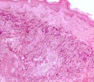
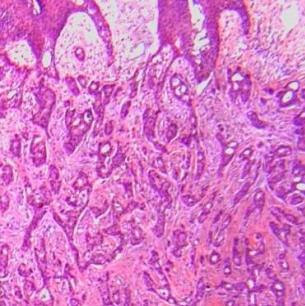
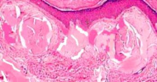
Answer of Dermatopathology Case 17

Fordyce's Spot (Ectopic sebaceous glands) Visit: Fordyce's Spot ; Visit: Dermatopathology site Abstract: Treatment of Fordyce spots with CO2 laser.Dermatol Surg. 2003 Aug;29(8):869-71. BACKGROUND: Fordyce spots are heterotopic sebaceous glands that can be located at the lips' vermilion or the oral mucosa. Although this is considered a rathercommon disorder, a treatment for this condition that sometimes affects patientsfrom only a cosmetic viewpoint has not yet been described. OBJECTIVE: To evaluateCO2 superpulsed laser treatment in two subjects with Fordyce spots. METHODS: Two patients with papules and yellowish plaques at the upper lip corresponding to Fordyce spots were treated with coherent Ambulase CO2 superpulsed laser (CoherentMedical, Palo Alto, CA) ; after informed consent was obtained, two to three passeswere performed in one session using 2 and 4 W and a spot size of 2 mm. RESULTS:Complete re-epithelization was observed 2 weeks later with no residual Fordyce papu

Dermatopathology Case 17

Image
Image1 Image2 Image3 Case 17 A 58 year old male with clusters of yellowish spots (2 mm in diameter) on the upper lip. Biopsy of the yellowish spot. Answer

Answer of Dermatopathology Case 16

Trichilemmoma Visit: Pathology of Trichilemmoma Visit: Dermatopathology site Abstract: Atypical clinical appearance and localization of trichilemmoma. a case report. Pathologica. 2009 Jun;101(3):133-4. Trichilemmoma is a benign cutaneous tumor that shows characteristics of differentiation similar to the outer hair sheath. We report the case of a woman presenting with a nodular tender mass of the back that was diagnosed as anisolated trichilemmoma. Several lines of evidence suggest that trichilemmoma should be considered in the differential diagnosis of any indistinct facialpapule. This report documents a non-facial example of trichilemmoma. Atypical clinical appearance and localization of this neoplasm in our patient suggest thatonly histological findings are specific of this tumor. Detection of Human Papillomavirus infection in trichilemmomas and verrucae using in situ hybridization. J Cutan Pathol. 2009 Background: It is hypothesized that trichilemmomas are 'burned out' verru

Dermatopathology Case 16

Image
Image 1 Image2 Image3 Image4 Case 16 A solitary, dome-shaped flesh coloured papule on the face of a 50 year old female. Answer

Answer of Dermatopathology Case 15

Cylindroma Visit: Pathology of Cylindroma Visit: Dermatopathology site Abstract: Cylindroma of the breast: a case report and review of the literature. Diagn Pathol. 2009 Sep 2;4:30. ABSTRACT: Cylindroma of the breast is a very rare lesion which is morphologicaland immunophenotypically identical to benign dermal cylindroma. We report a breast cylindroma in a previously healthy 62 year old female detected through anational breast screening program. The patient had no significant family or past medical history, and specifically no history of breast or skin diseases. The tumor consisted of well circumscribed islands of epithelial cells surrounded by adense membrane material, and focally containing hyaline globules. At low power the islands of tumour cells formed a "jig-saw" pattern, which is typical of cylindroma, but was present within normal breast parenchyma and no had directconnection with the overlying skin. Two distinct cell populations, smaller peripheral basaloid cells an

Dermatopathology Case 15

Image
Image 1 Image 2 Image3 Case 15 A nodule on the scalp of a 28 year old male. Answer

Answer of Dermatopathology Case 14

Warty Dyskeratoma Visit: Pathology of Warty Dyskeratoma Visit: Dermatopathology site Abstract: Multiple warty dyskeratomas on the scalp. Dermatol Online J. 2009 Feb 15;15(2):8. Warty dyskeratoma is usually characterized by solitary papules or nodules which reveal acantholytic dyskeratosis histopathologically. Warty dyskeratoma most commonly presents as a single lesion, but there are a few case reports of patients with multiple lesions. Herein we report a female patient presenting with multiple verrucous papules on the scalp diagnosed clinically and histopathologically as warty dyskeratoma. Warty dyskeratoma: infundibular histogenesis. Anatomoclinical study of 43 cases. Ann Dermatol Venereol. 2007 Aug-Sep;134(8-9):633-6. BACKGROUND: The exact origin and classification of warty dyskeratoma inepithelial tumours are still debated. The purpose of this study was to examinethe relationship between this tumour and the pilosebaceous follicles. MATERIALS AND METHODS: This was a retrospective, a

Dermatopathology Case 14

Image
Image1 Image2 Image3 Image4 Image5 Case 14 A greyish hyperkeratotic nodule with an umbilicated center, on the scalp of a 51 year old man. The nodule is 2 cm in diameter. Answer

Answer of Dermatopathology Case 13

Trichofolliculoma Visit: Pathology of Trichofolliculoma Visit: Dermatopathology site Abstract: A Revaluation of Trichofolliculoma: The Histopathological and Immunohistochemical Features. Am J Dermatopathol. 2009 Sep 1. Few investigations on the histopathology of trichofolliculoma (TF) have so far included an immunohistochemical study. To seek new insight into TF with are valuation of the histopathological features and an investigation of the immunohistochemical profile, 14 TFs were revaluated for the histopathology andthe immunohistochemical profile of various cytokeratins (CKs), hair follicle stem cell markers, and others. The CK15 expression was upregulated in the basal cells from the primary cystic structures beyond to secondary follicles without expression of CK19. CK16 and CK17 were positive in the suprabasal cells of the primary cystic structures and the immature secondary hair follicles. No exact isthmus/bulge region was seen in the anagen secondary hair follicles, and newly dev

Dermatopathology Case 13

Image
Image1 Image2 Image3 Case 13 A solitary flesh-coloured nodule on the face. Answer

Answer of Dermatopathology Case 12

Trichoepithelioma Visit: Pathology of trichoepithelioma Visit: Dermatopathology site Abstracts: A novel missense mutation of CYLD gene in a Chinese family with multiple familial trichoepithelioma. Arch Dermatol Res. 2009 Multiple familial trichoepithelioma (MFT, OMIM 601606) is an autosomal dominantly inherited disease. It is characterized by numerous skin-colored papules on the central face. Pathogenic mutations in the CYLD gene have been identified. In this report, we identified a novel mutation of CYLD gene in a Chinese family with MFT. It is a novel heterozygous nucleotide G-->A transition at position 2,317 in exon 17 of the CYLD gene. Our study expands the database on the CYLD gene mutations in MFT. Trichoepithelioma of the vulva: report of a case and review of the literature.J Low Genit Tract Dis. 2009 Trichoepithelioma is a rare benign skin lesion arising from hair follicles. It most commonly arises on the face and scalp. It is exceptionally rare on the vulva. We present a ca

Dermatopathology Case 12

Image
Image1 Image2 Image3 Image4 Case 12 A 25 year old male with a solitary tan coloured nodule on the right side of the face. Answer

Answer of Dermatopathology Case 11

Aggressive Digital Papillary Adenocarcinoma Visit: Pathology of Aggressive Digital Papillary Adenocarcinoma Visit: Dermatopathology site Abstract: Aggressive digital papillary adenocarcinoma : a review.Clin Exp Dermatol. 2009 Oct 23. Summary Vigorous treatment of aggressive digital papillary adenocarcinoma (ADPA), including amputation, has been recommended by most authors, but then appropriateness and effectiveness of excision as an alternative to amputation has not been systematically evaluated. To evaluate the appropriateness and effectiveness of excision as an alternative to amputation in the treatment of ADPA, we reviewed the clinical presentations, treatments and patient outcomespresented in case reports on ADPA available on Ovid MEDLINE. We also assessed theresults of immunohistochemical staining for proliferation markers in one patient in order to explain the nonaggressive nature of ADPA noted in that patient. Except for the duration of lesions, there was no significant differ

Dermatopathology Case 11

Image
Image1 Image2 Image3 Image4 Case 11 A 52 year old male with an enlarging nodule on the middle finger of the right hand. The lesion is 2 cm in diameter. Answer

Answer of Dermatopathology Case 10

Primary Cutaneous Actinomycosis Visit: Pathology of Actinomycosis Visit: Dermatopathology site Abstract: Cutaneous actinomycosis presenting as chronic mastitis. Clin Exp Dermatol. 2009 Apr 27. Summary Actinomycosis is a chronic granulomatous suppurative infection caused by anaerobic actinomyces. Primary cutaneous involvement is uncommon because of the exclusively endogenous habitat of the organism. We describe a very unusual presentation mimicking chronic mastitis. A 35-year-old woman presented 7 months post-partum with tenderness and induration in the right breast. She was pyrexial and felt systemically unwell. An initial diagnosis of mastitis was made.Treatment with penicillin, imipenem, co-amoxiclav and metronidazole had no effect. Skin biopsy revealed the characteristic 'sulphur granules' ofactinomycoses in the deep dermis. Long term oral clindamycin (> 12 months) has produced a very good response clinically, with a concomitant decrease ininflammatory markers. Cutaneous

Dermatopathology Case 10

Image
Image1 Image2 Image3 Image4 Image5 Case 10 A 25 year old female with an erythematous swelling, discharging pus and blood. The lesion is located on the right side of the neck and is present for last 2 years. Multiple small papules developed around the main lesion. Answer

Answer of Dermatopathology Case 9

Eccrine Spiradenoma Visit: Eccrine Spiradenoma Visit: Dermatopathology site Abstracts: Adenoid cystic carcinoma-like pattern in spiradenoma and spiradenocylindroma: a rare feature in sporadic neoplasms and those associated with Brooke-Spieglersyndrome.Am J Dermatopathol. 2009 Oct;31(7):642-8. Spiradenoma is a benign, morphologically well-defined cutaneous adnexal neoplasm that is closely related to cylindroma. We present the rare occurrence of adenoid cystic carcinoma (ACC)-like areas in 7 spiradenomas and 1 spiradenocylindroma, not described in the English literature to date. The ACC-like areas were a minor but significant component in all lesions and were usually multifocal and blended with the conventionally appearing parts of the neoplasms. The ACC-like areas weretypified by cribriform formations of epithelial cells concentrically arranged around gland-like spaces filled with mucin, homogeneous eosinophilic material, or granular basophilic material. In some neoplasms, only mucin oc

Dermatopathology Case 9

Image
Image1 Image2 Image3 Image4 Case 9 A firm painful nodule on the right forearm of a 35 year old male. Answer

Answer of Dermatopathology Case 8

Cutaneous Lymphadenoma Visit: Cutaneous lymphadenoma Visit: Dermatopathology site Abstracts: Glandular congenital lymphadenoma. Pediatr Dermatol. 2007 Sep-Oct;24(5):547-50. Cutaneous lymphadenoma is known to occur over a broad age range, from 14 to 72 years of age. We report the unique clinical and histologic presentation of acutaneous lymphadenoma from the suprapubic abdomen of a neonate which may represent a novel subtype, glandular congenital lymphadenoma. Cutaneous lymphadenoma is a rare tumor with a distinct histologic triad of epithelial nodules, dense fibrous stroma, and intense intranodular lymphocytic infiltrate.Typically, it is a slow growing, skin colored papule, nodule, or plaque,clinically resembling a basal cell carcinoma and often occurring in the head and neck region or lower extremities. Cutaneous lymphadenoma. J Am Acad Dermatol. 2003 Dec;49(6):1115-6 . Cutaneous lymphadenoma is a rare tumor with distinctive histologic features. This entity was originally described a

Dermatopathology Case 8

Image
Image1 Image2 Image3 Image4 Image5 Case 8 A dome-shaped, flesh coloured solitary nodule on the forehead of a 46 year old male. Answer

Answer of Dermatopathology Case 7

Primary Cutaneous Adenoid Cystic Carcinoma Visit: Cutaneous Adenoid Cystic Carcinoma Visit: Dermatopathology site Abstract: Primary cutaneous adenoid cystic carcinoma. J Am Acad Dermatol. 2008 Apr;58(4):636-41. Primary cutaneous adenoid cystic carcinoma is a rare, slow-growing malignancy first described by Boggio in 1975. This tumor characteristically consists of basophilic cells with a distinct adenoid or cribriform pattern in the mid to deep reticular dermis. Modified myoepithelial cells with prominent basement membrane material often surround true lumina. Definitive diagnosis relies on the characteristic histologic features and the exclusion of metastatic disease. We describe two patients who presented with painful papules of the scalp and weresuccessfully treated with wide local excision. Adenoid cystic carcinoma. Actas Dermosifiliogr. 2006 Nov;97(9):578-80. Adenoid cystic carcinoma is an uncommon tumor of the head and neck. Although itis mainly located in the salivary gland, a s

Dermatopathology Case 7

Image
Image1 Image2 Image3 Image4 Image5 Case 7 A 58 year old male with an ulcerated nodule on the scalp. ? BCC Answer

Answer of Dermatopathology Case 6

Primary Cutaneous Apocrine Carcinoma Visit: Cutaneous Apocrine Carcinoma Visit: Dermatopathology site Abstract Related article Primary cutaneous cribriform apocrine carcinoma: a clinicopathologic andimmunohistochemical study of 26 cases of an under-recognized cutaneous adnexal neoplasm. J Am Acad Dermatol. 2009 Oct;61(4):644-51. BACKGROUND: Cribriform carcinoma is the histopathologic variant of cutaneousapocrine carcinoma characterized by interconnected solid aggregations of neoplastic cells that are punctuated by small round spaces. OBJECTIVE: To describe the histopathologic and immunohistochemical characteristics of this under-recognized cutaneous adnexal neoplasm. METHODS: Twenty-six cases of primarycutaneous cribriform apocrine carcinoma were clinically, histopathologically, and immunohistochemically studied. RESULTS: Seven neoplasms arose in males and 19 in females. The median age of the patients was 47.8 years. The lower and upper limbswere the most frequent sites. Histopathologi

Dermatopathology Case 6

Image
Image1 Image2 Image3 Image4 Case 6 A 68 year old man with a lump in the left axilla, 4 cm in diameter. Answer

Answer of Dermatopathology Case 5

Colloid Milium Visit: Colloid Milium Visit: Dermatopathology site Abstract: Colloid milium: a review and update. J Drugs Dermatol. 2007 Mar;6(3):293-6. Colloid milium (CM) is a rare cutaneous deposition disease with at least 3 distinct subtypes. The exact histogenesis of the condition is still unresolvedand awaits definitive elucidation. Electron microscopy and immunohistochemicalanalysis have allowed the distinction of CM from clinically similar conditionssuch as amyloidosis. Successful treatment has been achieved with dermabrasionand, more recently, with ablative and fractional laser resurfacing of affected skin. Colloid milium: a rare cutaneous deposition disease. J Pak Med Assoc. 2008 Apr;58(4):207-9. Colloid milium is a rare degenerative skin disorder known by the development of small translucent, yellowish brown papular nodules or plaques, generally located in sun exposed areas. Clinically they are of two types, adult and juvenile type. We present a case of adult type Colloid mil

Dermatopathology Case 5

Image
Image1 Image2 Case 5 A 75 year old male with a flesh coloured papule on the right side of the face. Answer