Posts

Answer of Dermatopathology Case 4

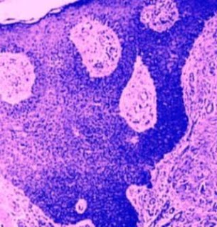
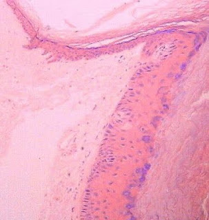
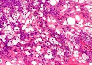
Eccrine Porocarcinoma Visit: Eccrine Porocarcinoma Visit: Dermatopathology site Abstract: Eccrine porocarcinoma of the auricle: a case report. Kaohsiung J Med Sci. 2009 Jul;25(7):401-4. Eccrine porocarcinoma (EP) is a rare skin malignant lesion representing0.005-0.01% of all cutaneous tumors. It is a tumor that most commonly present in elderly people aged over 60 years. Approximately 250 cases of EP have been reported since this disease was first described in 1963. However, only three cases occurring specifically on the ear (including the current case) have beendocumented in the literature to date. Based on the rarity of EP of the ear, we present this 78-year-old man with EP on the right ear lobule, which was diagnosedaccidentally during the management of other unrelated problems. The etiology,diagnosis, treatment and prognosis of this disease are discussed, with a brief review of the literature in this report. Fine-needle aspiration cytology of metastatic eccrine porocarcinoma.Diagn

Dermatopathology Case 4

Image
Image1 Image2 Image3 Image4 Image5 Image6 Case 4 A 70 year old female with an ulcerated, cauliflower like plaque on the left thigh. ? SCC Answer

Answer of Dermatopathology Case 3

Large Cell Acanthoma Visit: Large cell Acanthoma Visit: Dermatopathology site Abstracts: A conjunctival lesion with histological features similar to large-cell acanthoma of the skin.J Cutan Pathol. 2009 Aug 27. Large-cell acanthoma (LCA) is a sharply demarcated epidermal lesion composed oflarge keratinocytes and characterized by hyperkeratosis, hypergranulosis and orthokeratosis. It usually affects actinically damaged skin. LCA is currentlybelieved to be a distinct entity with an unclear pathogenesis and a benign biologic behavior. Here, we describe the first occurrence of LCA in theconjunctiva. The lesion recurred following initial excision, and later recurred as a carcinoma in situ following a second excision. This case suggests that LCA may have a malignant and transformation potential. Ghazi NG, Patel BS, OlsakovskyLeslie A, White K, Patterson JW. A conjunctival lesion with histological features similar to large-cell acanthoma of the skin. Large cell acanthoma.Int J Dermatol . 2003

Dermatopathology Case 3

Image
Image1 Image2 Image3 Image4 Case 3 A solitary tan brown macule on the left upper upper arm of a 45 year old woman. The lesion is 10mm in diameter. Answer

Answer of Dermatopathology Case 2

Sebaceous adenoma in a patient with Muir-Torre syndrome Visit: Dermatopathology site Visit: Sebaceous Adenoma Abstract: Cutaneous sebaceous neoplasms as markers of Muir-Torre syndrome: a diagnosticalgorithm. J Cutan Pathol. 2009 Jun;36(6):613-9. Sebaceous gland neoplasms such as adenoma, epithelioma, and carcinoma areuncommon cutaneous tumors. Although sporadic, their occurrence is clinicallysignificant because of their association with Muir-Torre syndrome (MTS). MTS is a rare autosomal dominant genodermatosis characterized by the occurrence of sebaceous gland neoplasms and/or keratoacanthomas associated with visceralmalignancies that include gastrointestinal and genitourinary cancers. MTS isusually the result of germline mutation in one or more of the DNA mismatch repair(MMR) genes. MMR genes commonly implicated include MSH-2 and MLH-1 and, morerecently, MSH-6. Recent evidence suggests that immunohistochemistry is verysensitive and effective in detecting these defects in cutaneous tum

Dermatopathology Case 2

Image
Image1 Image2 Image3 Image4 Image5 Case 2 A 71 year old male with multiple yellowish nodule on the scalp. Patient has a history of adenocarcinoma of the colon. These are sections from one of the yellowish nodule. Answer

Answer of Dermatopathology Case 1

Cutaneous Leiomyosarcoma Visit: Leiomyosarcoma Visit: Dermatopathology site Abstracts: Cutaneous leiomyosarcoma originating in a symplastic pilar leiomyoma: a rareoccurrence and potential diagnostic pitfall.J Cutan Pathol. 2009 Sep 22. A case of cutaneous leiomyosarcoma originating in a symplastic pilar leiomyoma isdescribed. The patient was a 60-year-old man who presented with a painless noduleon his chest. Thorough clinical examination did not reveal any evidence of tumor elsewhere. A punch biopsy was performed which showed a benign spindle cellneoplasm with focal cellular pleomorphism that had smooth muscle differentiation and was clearly originating from the arrector pili muscle, consistent with acutaneous symplastic pilar leiomyoma. Immunohistochemical studies were performed and the tumor cells were strongly positive for smooth muscle actin (SMA) anddesmin. Given the clinical information of a mass on the chest, clinicalevaluation was recommended and a re-excision was performed. H

Dermatopathology Case 1

Image
Image1 Image2 Image3 Image4 Image5 Image6 Case-1 A 65 year old female with a skin nodule 0.9cm in diameter, on the right upper arm. Smooth muscle actin and desmin : Positive. Cytokeratin, EMA, S100 protein: Negative Answer