Posts

Answer of Dermatopathology Case 52

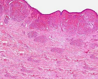
Acquired Tufted Angioma Visit: Dermatopathology site Visit: Pathology of Acquired Tufted Angioma Abstract: Perianal acquired tufted angioma associated with pregnancy: case report. Tech Coloproctol. 2002 Sep;6(2):117-9. Tufted angiomas are rare lesions described as slowly growing/spreading erythematous macules especially located in the upper trunk and neck. Herein we report the case of perianal location of a tufted angioma in a young pregnant woman. She came to our observation complaining of perianal pain accompanied by bleeding at defecation. A lesion resembling a perianal fissure was observed. Mild hypertonia of the internal sphincter was confirmed at manometry. After one week of ineffective medical treatment, surgery was planed at the end of the sixteenth week under local anaesthesia. The lesion was excised and a minimal sphincterotomy was performed; histopathology report described features of a tufted angioma. The pregnancy proceeded regularly, without anal symptoms, followed by nor

Dermatopathology Case 52

Image
Image1 Image2 Image3 Case 52 A 10 year old boy with a raised papule on the neck. ? Pyogenic granuloma. Answer

Answer of Dermatopathology Case 51

Prurigo Nodularis Visit: Dermatopathology site Visit: Pathology of Prurigo Nodularis Abstract: Prurigo nodularis: systematic analysis of 58 histological criteria in 136 patients.J Cutan Pathol. 2010 May;37(5):578-86. Epub 2009 Nov 30. BACKGROUND: To date, there has been no systematic investigation of the detailed histological features of prurigo nodularis (PN) in a large cohort of patients. METHODS: This retrospective study includes skin biopsies of 136 patients (63 males, 73 females; mean age: 58.38 years) with PN. RESULTS: Highly characteristic for PN is the presence of thick compact orthohyperkeratosis; the hairy palm sign (folliculosebaceous units in nonvolar skin in conjunction with a thick and compact cornified layer, like that of volar skin); irregular epidermal hyperplasia or pseudoepitheliomatous hyperplasia; focal parakeratosis; hypergranulosis; fibrosis of the papillary dermis with vertically arranged collagen fibers; increased number of fibroblasts and capillaries; a superf

Dermatopathology Case 51

Image
Image1 Image2 Case 51 A 35 year old male, with a few intensely pruritic, firm pink nodules on the extensor aspect of the left upper arm. Answer

Answer of Dermatopathology Case 50

Cutaneous Herpes Simplex Infection Visit: Dermatopathology site Visit: Herpes Simplex Infection Abstract: Histopathology of the more common viral skin infections. Actas Dermosifiliogr .2010 Apr;101(3):201-16. We describe the histopathological characteristics of viral skin infections. Herpes simplex virus and varicella-zoster virus produce an intraepidermal vesicle with variable degrees of epithelial necrosis. Typical findings include keratinocytes with ballooned nuclei with a ground-glass appearance and giant multinucleated keratinocytes. In the endothelial cells of the dermal blood vessels, cytomegalovirus produces large eosinophilic nuclear inclusions surrounded by a clear halo. Human herpes virus 8 is etiologically associated with Kaposi sarcoma. In its early stages, this tumor contains blood vessels with a fine endothelium passing through the dermal collagen bundles. In the plaque and nodular stages, the vessel lumens are more clearly visible and there is a progressive increase in