Posts

Showing posts from January, 2010

Answer of Dermatopathology Case 37

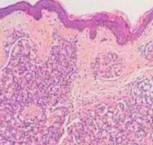
Dermatitis Herpetiformis Visit: Dermatopathology site Abstract: Dermatitis herpetiformis. An update of the pathogenesis.Hautarzt. 2009 Aug;60(8):627-30, 632. The multifactorial pathogenesis of dermatitis herpetiformis is reviewed in light of current experimental data. Genetic background, gluten consumption, and abnormal immune and autoimmune reactions are the most important pathogenetic factors, but other agents also participate in the disease development. The predisposing and inducing factors are summarized, while the pathophysiological steps leading to the development of skin symptoms are detailed. Dermatitis herpetiformis. A clinical chameleon.Hautarzt. 2006 Nov;57(11):1021-8; quiz 1029. Celiac disease is a genetically determined bowel disease also influenced by exogenous factors in which exposure to grain components triggers a chronic immune response with intestinal symptoms. Dermatitis herpetiformis represents the cutaneous manifestation of celiac disease. While intense pruri...

Dermatopathology Case 37

Image
Image1 Image2 Image3 Case 37 A 35 year old man with small intensely itchy blisters and papules on both the elbows. The patient has a history of celiac disease. Answer

Answer of Dermatopathology Case 36

Angiolymphoid Hyperplasia with Eosinophilia (Epithelioid Hemangioma) Visit: Angiolymphoid hyperplasia with eosinophilia(Epithelioid Hemangioma) Visit: Dermatopathology site Visit: Pathology of Vascular Tumours Abstract: Epithelioid hemangioma (angiolymphoid hyperplasia with eosinophilia) of theorbit: a case report. J Med Case Reports. 2007 Jun 25;1:30. ABSTRACT: BACKGROUND: Angiolymphoid hyperplasia with eosinophilia (ALHE) and Kimura's Disease (KD) share many clinical and histopathological features. Although they were once considered different stages of the same disease, they arenow known to represent separate entities. Recently, ALHE is being called epithelioid hemangioma (EH), a term that better describes the possible neoplastic nature of the entity. CASE PRESENTATION: An eighteen year-old Asian female presented with a three-month history of fluctuating swelling and ptosis of the left upper eyelid. Computed tomography disclosed a distinct homogeneous lesion in the left superior...

Dermatopathology Case 36

Image
Image 1 Image 2 Image 3 Image 4 Case 36 A 32 year old Asian man presenting with a dome-shaped, crusted nodule (3 cm in diameter) on the scalp. Answer

Answer of Dermatopathology Case 35

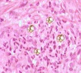
Chromomycosis (Chromoblastomycosis) Visit: Pathology of Chromoblastomycosis(Chromomycosis) ; Visit: Dermatopathology site ; Visit: Infectious Disease Online . Abstract: Difficult mycoses of the skin: advances in the epidemiology and management of eumycetoma, phaeohyphomycosis and chromoblastomycosis.Curr Opin Infect Dis. 2009 Dec;22(6):559-63. PURPOSE OF REVIEW: Eumycetoma, phaeohyphomycosis and chromoblastomycosis are subcutaneous mycoses having in common the fact that they are acquired as a result of penetrating trauma to the skin and may be caused by a large variety of fungi. This article will review recent data regarding the epidemiology and treatment of these infections. RECENT FINDINGS: Recent epidemiologic observations in these mycoses include an increased incidence of phaeohyphomycosis in immunosuppressed patients, the association of polymorphisms in genes involved in innate immunity, the occurrence of eumycetoma caused by Madurella mycetomatis and the nosocomial acquisition of...

Dermatopathology Case 35

Image
Image 1 Image 2 Image 3 Image 4 Case 35 A 42 year male with a verrucous, warty plaque in the distal part of the left extremity. Answer

Answer of Dermatopathology Case 34

Bacillary Angiomatosis Visit: Pathology of bacillary angiomatosis Visit: Dermatopathology site Visit: Pathology of Vascular Tumours Abstracts: Bartonellosis. Clin Dermatol. 2009 May-Jun;27(3):271-80. Bartonella spp are fastidious bacteria that occur in the blood of man and mammals; they are usually vector borne but can also be transmitted by animal scratches and bites. The bartonelloses of medical importance comprise Carrión's disease, trench fever, cat-scratch disease, bacillary angiomatosis, and peliosis hepatis. Carrión's disease, known as Oroya fever in the acute phase and verruga peruana (Peruvian wart) in its chronic form, has curious manifestations that,until recently, have been restricted in their geographic distribution to dwellers of the high, dry Andean valleys, but new sites of disease are emerging. Trench fever is associated with louse-borne disease and homelessness. Cat-scratch disease, bacillary angiomatosis, and peliosis hepatis are increasingly being recognize...

Dermatopathology Case 34

Image
Image1 Image2 Case 34 A 36 year old HIV- positive woman presenting with multiple red papules all over her body. Answer

Answer of Dermatopathology Case 33

Glomeruloid Hemangioma Visit: Glomeruloid Hemangioma Visit: Dermatopathology site Visit: Pathology of Vascular Tumours Abstract: POEMS syndrome with an isolated lytic lesion and glomeruloid hemangiomas: report of one case. Rev Med Chil. 2009 May;137(5):680-4. Epub 2009 Jul 17. POEMS syndrome (polyneuropathy, organomegaly, endocrine abnormality, M-protein, plasma cell dyscrasia, and skin lesions) is a rare atypical plasma cell dyscrasia with characteristic para neoplastic manifestations. Glomeruloid hemangioma is a typical skin change pathogenetically related with elevated levels of Vascular Endothelial Growth Factor (VEGF). We report a 69 year-old woman that presented cachexia associated with diabetes, hypothyroidism and severe sensitive motor polyneuropathy. Her skin changes included hyper pigmentation, acrocyanosis and glomeruloid hemangioma. The subsequent study revealed a monoclonal gammopathy lambda type; a unique lytic vertebral lesion and a clonal plasma cell proliferation. Trea...

Dermatopathology Case 33

Image
Image1 Image2 Image3 Image4 Case 33 A female 55 year old with a history of Poem's syndrome. Multiple violaceous papules (5 mm in diameter) are present on the left upper limb. Answer

Answer of Dermatopathology Case 32

Sebaceous Hyperplasia Visit: Dermatopathology site ; Visit: Sebaceous Hyperplasia Abstract: Sebaceous gland hyperplasia of the foreskin.Clin Exp Dermatol. 2009 Apr;34(3):372-4. Epub 2008 Dec 9. Two men, aged in their 20s, presented with multiple, soft, rounded papules on the prepuce. The lesions were centrally umbilicated, resembling molluscum contagiosum, but clearly distinct from Tyson's glands. Surface microscopy showed well-defined, milky-white, bag-shaped structures, which under histological examination were found to be sebaceous glands with various features of hyperplasia. A lymphocytic T-cell infiltrate, closely associated with progressive degeneration and destruction of the sebocytes, was visible around the glands. In the differential diagnosis of penile papular lesions, this unusual clinicalnpresentation supported by dermatoscopy is consistent with preputial sebaceous gland hyperplasia. As both patients had a prominent T-cell infiltration, it ispossible that under inflamma...

Dermatopathology Case 32

Image
Image1 Image 2 Case 32 A 45 year old female with a solitary yellowish, umbilicated papule 2.5mm in diameter. The lesion is located on the left side of the face. Answer

Answer of Dermatopathology Case 31

Reticulohistiocytoma Visit: Dermatopathology site ; Abstract: Reticulohistiocytoma (solitary epithelioid histiocytoma): a clinicopathologic and immunohistochemical study of 44 cases. Am J Surg Pathol. 2006 Apr;30(4):521-8. Reticulohistiocytoma and multicentric reticulohistiocytosis are designations for uncommon, incompletely characterized histiocytic proliferations of the skin or soft tissues. In this study, we analyzed a uniform group of44 lesions composed of epithelioid histiocytes, comprising a subset of lesions originally designated as reticulohistiocytoma, and propose designating them as "solitary epithelioid histiocytoma" (SEH), in line with the recently published classification proposal for histiocytic disorders. There were 26 males and 18 females with a median ageof 35 years (range, 2.5-74 years). All patients had a superficial, circumscribed, mildly elevated, solitary lesion (size range, 1.5-11 mm; median, 4 mm), located in the trunk wall (n = 16), lower extremity ...

Dermatopathology Case 31

Image
Image1 Image2 Image3 Case 31 A 38 year old female with a superficial, circumscribed, mildly elevated, solitary lesion. The lesion is 5 mm in diameter and located on the forehead. Answer

Answer of Dermatopathology Case 30

Xanthelasma Visit: Xanthelasma ; Visit: Dermatopathology site ; Visit: Verruciform Xanthoma ; Visit: Gastric Xanthoma Visit: Eye Pathology Online . Abstract: Xanthelasma and juvenile xanthogranuloma in a 7-year-old boy. Ann Dermatol Venereol. 2009 Oct;136(10):723-6. Epub 2009 May 15. BACKGROUND: Palpebrum xanthelasma is the most common type of xanthoma seen inadults but it is extremely rare in children. We report an original case of bilateral xanthelasma palpebrarum associated with juvenile xanthogranuloma (JXG) in a 7-year-old child. Only two cases of xanthelasma in children have been described to date. The association of xanthelasma and JXG has never been described. PATIENTS AND METHODS: A 7-year-old boy presented xanthelasmas on both eyelids. At the same time, pinkish JXG papules appeared on the child's trunk. The boy had been diagnosed at the age of 10 months with myelogenous leukaemia, which was in remission. He also had a familial history of hypercholesterolaemia. The ski...

Dermatopathology Case 30

Image
Image1 Image2 Image3 Case 30 A 40 year old female with a yellow, soft plaque on the upper eyelid. Answer

Answer of Dermatopathology Case 29

Pilomatrix Carcinoma Visit: Pathology of Pilomatrix Carcinoma Visit: Dermatopathology site Abstract: Pilomatrix carcinoma presenting as an extra axial mass: clinicopathological features. Diagn Pathol. 2008 Nov 29;3:47. ABSTRACT: Pilomatrix carcinoma is the rare malignant counterpart of pilomatrixoma, a skin adnexal tumour originating from hair matrix cells. Pilomatrix carcinoma can arise as a solitary lesion de novo, or through transformation of a pilomatrixoma. Pilomatrixoma was first described erroneously as being of sebaceous gland origin but was later discovered to be derived from hair matrix cells. They are rare, slow growing tumours of the skin found in thelower dermis and subcutaneous fat and are predominantly found in the neck and the scalp. While known to be locally aggressive, no malignant form was thought toexist until it was described relatively recently. Since then, approximately ninety cases of pilomatrix carcinoma have been reported.We report the case of a 41 year old me...

Dermatopathology Case 29

Image
Image1 Image2 Image3 Image4 Case 29 A 56 year old male with a skin lump, 5 cm in diameter, on the posterior part of the neck. Answer (Compare with Case Number 23)