Posts

Answer of Dermatopathology Case 108

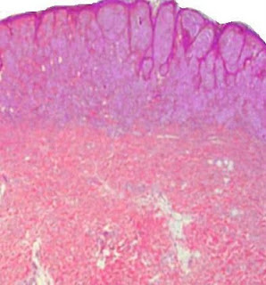
Median Raphe Cyst Visit: Dermatopathology Site Visit: Pathology of Median Raphe Cyst Abstract: Median raphe cyst of the penis.Dermatol Online J. 2005 Dec 1;11(3):37. Cysts of the median raphe are uncommon. We describe a 43-year-old man with an asymptomatic nodule on the glans penis. Excision of the lesion was performed followed by histological and immunohistochemical studies. Histopathological examination revealed a solitary unilocular cystic cavity in the corium, lined by a pseudostratified columnar epithelium, among which mucinous cells where identified. Hints of decapitation secretion occurred at the apex of the luminal layer. An immunohistochemical study using a standard avidin-biotin peroxidase method disclosed CK7 and CK13 reactivity in the columnar cells. Epithelial membrane antigen (EMA) and carcinoembryonic (CEA) antigen immunoreactivity occurred at the apical border of the luminal cells. No staining was obtained with anti-CK20, human milk fat globulin 1 (HMFG1) and anti- S10

Dermatopathology Case 108

Image
Image1 Image 2 Image3 Image4 Case 108 A 38 year old male with a nodule on the glans penis. Diagnosis

Answer of Dermatopathology Case 107

Naevoid Melanoma Visit: Dermatopathology Site Visit: Pathology of Naevoid Melanoma In general, common naevi are diagnosable at low power. It is important to exclude the possibility of naevoid melanoma. This is a rare variant that mimics benign naevi and is difficult to recognise. The correct diagnosis is frequently made only after the patient has developed a metastasis. At low power, naevoid melanoma may have a nodular architecture, and exhibit other features of a common banal naevus, including circumscription and at least relative symmetry. Naevoid melanoma lacks the prominent junctional activity and pagetoid spread usually associated with superficial spreading melanoma. Common intradermal naevus “matures” (there is an overall decrease in nest size and cellular and nuclear size with depth). On low power examination, naevoid melanoma may appear to mature with depth, yet closer inspection reveals that the cells at the base of the lesions are similar in size to those of the superficial

Dermatopathology Case 107

Image
Image 1 Image 2 Image 3 Image 4 Image 5 Case 107 A 45 year old female with a pigmented lesion on the left side of the chest. Diagnosis

Answer of Dermatopathology Case 106

Desmoplastic Spitz Naevus Visit: Dermatopathology Site Visit: Pathology of Desmoplastic Spitz Naevus Visit: Pathology of Spitz Naevus Abstract: Desmoplastic Spitz nevus: a histopathological review and comparison with desmoplastic melanoma. Ann Dermatol Venereol. 2009 Oct;136(10):689-95. INTRODUCTION: Desmoplastic Spitz nevus is a rare variant of spitzoid tumours characterized by dermal proliferation of large epitheliod and/or fusiform melanocytes within a desmoplastic stroma, comprising thick, eosinophilic collagen bundles. It occurs most frequently in young adults and children, and exhibits a predilection for the limbs. Histologically, this entity may be mistaken for cutaneous fibrous tumours or desmoplastic melanoma. To establish useful histological criteria for differential diagnosis of desmoplastic Spitz nevus, we conducted a retrospective study comparing data concerning the clinical, demographic and histological characteristics of desmoplastic Spitz nevus and desmoplastic melanom